
Granulocyte Collection
Granulocytes are a crucial component of our immune system, tirelessly working to protect us from harmful invaders


All Professionally Made to Order for Quick Shipping
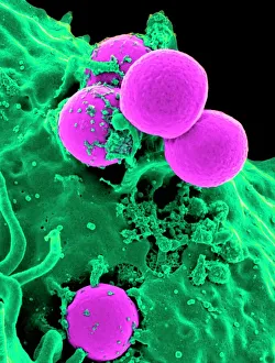
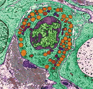
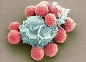
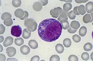
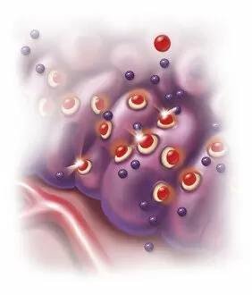
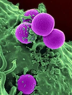
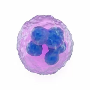
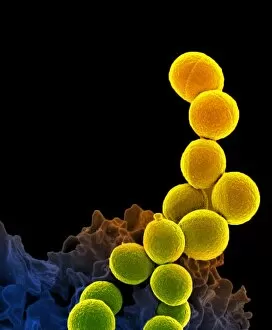
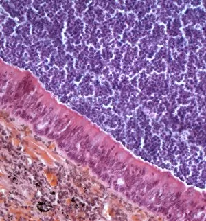
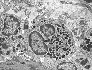
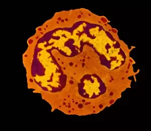
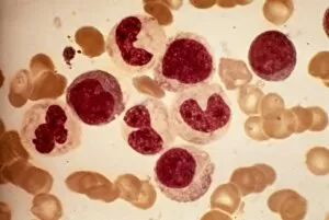
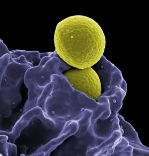
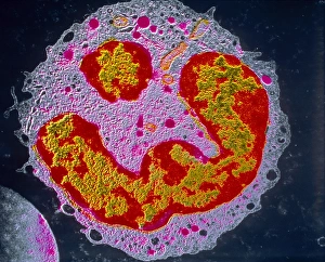
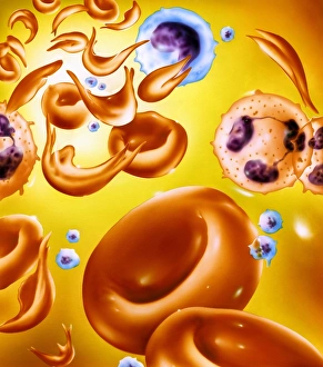
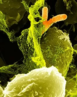
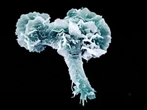
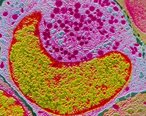
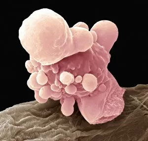
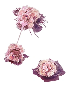
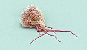
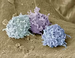
Granulocytes are a crucial component of our immune system, tirelessly working to protect us from harmful invaders. One such the neutrophil, depicted in a stunning scanning electron micrograph engulfing MRSA bacteria. Its relentless pursuit of pathogens showcases its effectiveness as a defender. Basophil white blood cells also play an essential role in our body's defense mechanism. A conceptual image portrays these unique cells, highlighting their importance in triggering allergic reactions by releasing histamine. Phagocytosis, the process by which granulocytes engulf and destroy foreign substances, is beautifully captured in another SEM image depicting fungal spores being devoured by these powerful defenders. Micrographs offer us glimpses into the intricate world of blood cell development. The myeloblast and promyelocyte stages are showcased through light microscopy images, showcasing the transformation from precursor cells to mature granulocytes. However, when surrounding tissue becomes inflamed due to infection or injury, several systemic effects can occur. This inflammation triggers various responses that can lead to discomfort and illness. To counteract allergic reactions caused by basophils' histamine release, antihistamines step in to block histamine receptors effectively. This prevents further allergic symptoms from occurring and provides relief for those affected. The complexity of granulocytes is further highlighted through artwork representing them as white blood cells with distinct features and functions. These illustrations help visualize their significance within our immune system. A blood smear under light microscopy offers yet another perspective on these vital defenders circulating within our bloodstream. The intricacies of their structure become apparent as they navigate through vessels ready to combat any threat encountered along the way. In some instances where infections persist or intensify, unfortunate outcomes may arise - illustrated by an SEM image showing MRSA bacteria alongside a dead neutrophil. This serves as a reminder that even though they are formidable warriors against invading pathogens.