
Guard Cell Collection
"Unlocking the Secrets of Guard Cells: A Closer Look at Leaf Pores" Delving into the microscopic world, we explore the intricate realm of guard cells


All Professionally Made to Order for Quick Shipping
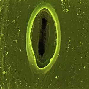
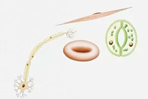
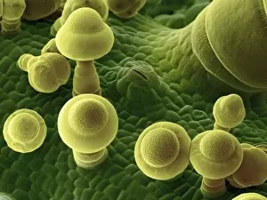
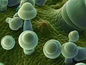
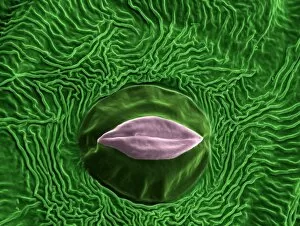
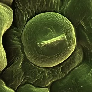
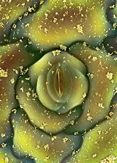
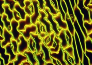
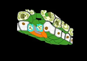
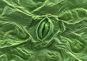
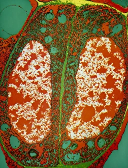
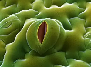
"Unlocking the Secrets of Guard Cells: A Closer Look at Leaf Pores" Delving into the microscopic world, we explore the intricate realm of guard cells. Through scanning electron microscopy (SEM), we witness nature's remarkable design in English oak leaf pores and French lavender leaf pores, revealing their unique structures. Intriguingly, chives leaf stoma captured under SEM C016/8060 showcases a mesmerizing pattern, while Coltsfoot leaf stomata (SEM C016/8052) exhibit an awe-inspiring arrangement. These images highlight the diversity and beauty found within plant anatomy. Illustrating various cell types alongside guard cells - nerve cells, red blood cells, muscle cells - emphasizes their vital role in sustaining life. The illustration serves as a reminder of the complexity that lies within every living organism. Further exploration takes us to tobacco leaves (SEM C016/9465) and squash leaves (SEM C016/9463), where guard cells stand out with their distinctive shapes and patterns. Thale cress leaves also reveal fascinating stoma formations under SEM C016/9460 and SEM C016/9459. As our journey continues through different plant species such as squash leaves (SEM C016/9464) and sorrel leaves (SEM C016 / 8059), we gain insight into how these plants regulate gas exchange through their specialized guard cells. These captivating images provide a glimpse into the hidden world of plant physiology. By studying guard cells using advanced imaging techniques like ESEM, scientists unravel secrets crucial for understanding how plants adapt to changing environments. Through this visual exploration, we appreciate nature's intricacy on a microscopic level while gaining admiration for the unsung heroes known as guard cells – guardians responsible for maintaining balance within every leaf's ecosystem.