
Helices Collection
"Unraveling the Mysteries of Helices: Exploring the Intricate World of Molecular Structures" DNA Transcription Unveiled


All Professionally Made to Order for Quick Shipping
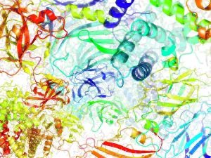
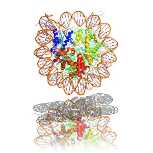
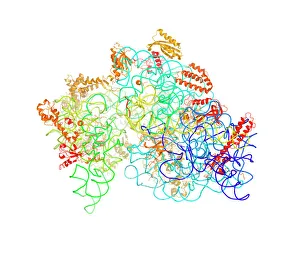
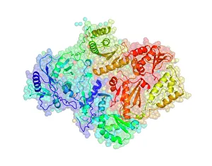
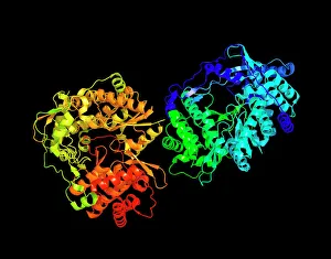
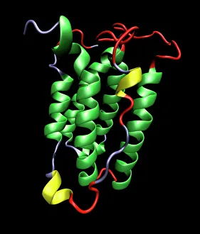
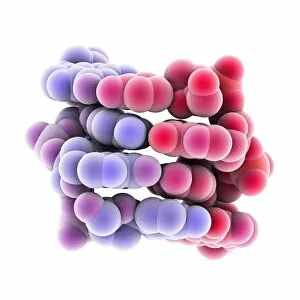
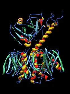
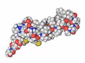
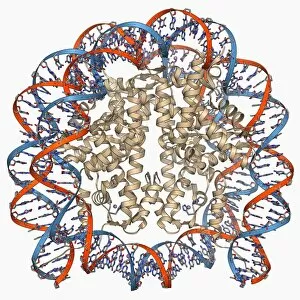
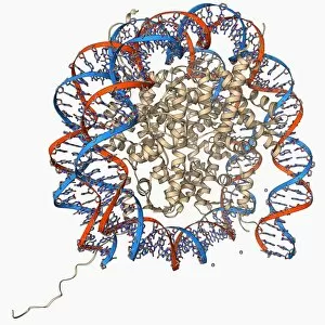
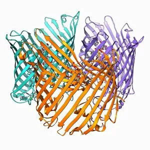
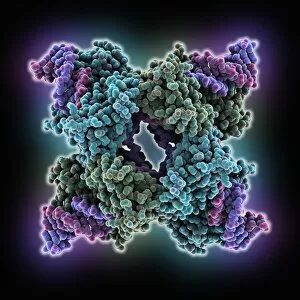
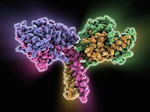
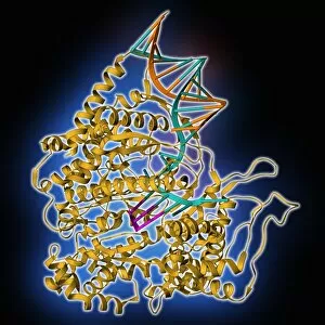
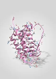
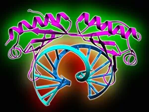
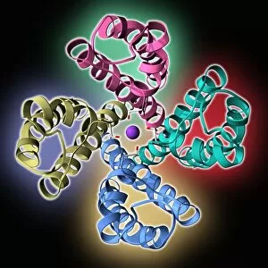
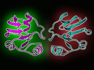
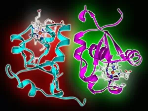
"Unraveling the Mysteries of Helices: Exploring the Intricate World of Molecular Structures" DNA Transcription Unveiled: Witness the intricate dance as DNA unravels and transcribes its genetic code, paving the way for life's blueprint. A Symphony in Proteins: Secondary structures adorned with helical motifs create a mesmerizing molecular model, showcasing nature's artistic prowess. Nucleosome Molecule - Nature's Genetic Sculptor: Behold the elegant architecture of nucleosomes, where DNA elegantly wraps around histone proteins forming a helical masterpiece. The Dance of Life: An artwork depicting a vibrant DNA molecule reveals its double-helix structure, symbolizing our very essence and genetic heritage. Bacterial Ribosome - The Protein Factory: Dive into the intricacies of bacterial ribosomes as they diligently translate genetic information into functional proteins through their complex helical framework. HIV Reverse Transcription Enzyme - Decoding Viral Secrets: Explore how this remarkable enzyme utilizes helical structures to reverse-transcribe viral RNA into infectious DNA, perpetuating HIV's stealthy invasion. Hepatitis C Virus Enzyme - Unmasking an Invisible Threat: Peer inside the molecular model of this cunning enzyme that hijacks human cells by employing intricate helices to replicate itself relentlessly. Interferon Molecule - Our Body's Defender: Discover how these mighty molecules employ their unique helical shape to activate our immune system against invading pathogens and protect us from harm. Z-DNA Tetramer Molecule C015/6557 – Unlocking New Dimensions in Genetics: Delve into the fascinating world of Z-DNA tetramers as they challenge conventional double-helical structures, offering new insights into gene regulation and potential therapeutic avenues. Cholera Toxin – A Deadly Ballet on a Molecular Stage.