Fraud Blocker Skip to main content

Histopathology Collection

Histopathology is a fascinating field that delves into the intricate details of diseases and their impact on our bodies

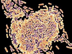
Background imageHistopathology Collection: Salmonella bacteria, SEM

Salmonella bacteria, SEM
Salmonella bacteria, coloured scanning electron micrograph (SEM). These Gram-negative rod-shaped bacteria can cause food poisoning when eaten in contaminated food

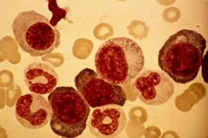
Background imageHistopathology Collection: Dohle bodies in blood cell, micrograph

Dohle bodies in blood cell, micrograph
Dohle bodies in blood cell. Light micrograph of a neutrophil white blood cell (centre) with Dohle bodies. These are small inclusions within the cells cytoplasm

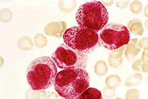
Background imageHistopathology Collection: Acute promyelocytic leukaemia, micrograph

Acute promyelocytic leukaemia, micrograph
Acute promyelocytic leukaemia. Light micrograph of blood cells from bone marrow in a case of acute promyelocytic leukaemia. Leukaemia is a cancer where certain blood cells form in excess

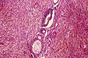
Background imageHistopathology Collection: Ovarian cancer, light micrograph C015 / 7103

Ovarian cancer, light micrograph C015 / 7103
Ovary cancer. Light micrograph of a section through a Krukenberg tumour (down centre) of the ovary. This is a secondary cancer that spreads (metastasises) from the digestive system

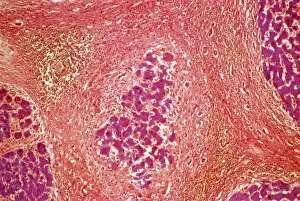
Background imageHistopathology Collection: Liver tissue cirrhosis, light micrograph

Liver tissue cirrhosis, light micrograph
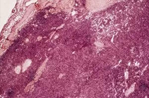
Liver cirrhosis. Light micrograph of a section through liver tissue showing cirrhosis. This is a condition where the liver responds to the injury or death of some of its cells by producing
Background imageHistopathology Collection: Cystic fibrosis

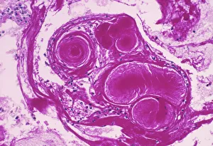
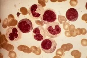
Cystic fibrosis. Light micrograph of a section through a bronchus (airway) of the lungs in a case of cystic fibrosis (mucoviscidosis)

Background imageHistopathology Collection: Osteoporotic bone

Osteoporotic bone

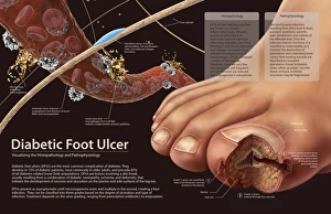
Background imageHistopathology Collection: Histopathology and pathophysiology of diabetic food ulcers

Histopathology and pathophysiology of diabetic food ulcers

Background imageHistopathology Collection: Gout crystals

Gout crystals. Polarised light micrograph of uric acid crystals in the synovial fluid (a lubricating liquid in joints) from a patient with gout

Background imageHistopathology Collection: Chronic lymphocytic leukaemia, micrograph

Chronic lymphocytic leukaemia, micrograph
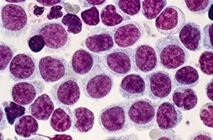
Chronic lymphocytic leukaemia. Light micrograph of blood cells sampled from a lymphatic ganglion in a case of chronic lymphocytic leukaemia, also called chronic lymphoid leukaemia (CLL)

Background imageHistopathology Collection: HIV particles, TEM

HIV particles, TEM
HIV (human immunodeficiency virus) particles, coloured transmission electron micrograph (TEM). Each particle consists of an RNA (ribonucleic acid, purple/brown) genome

Background imageHistopathology Collection: Lymphocytes in hair follicle, SEM

Lymphocytes in hair follicle, SEM
Lymphocytes in hair follicle, coloured scanning electron micrograph (SEM). Large numbers of lymphocytes (yellow) are seen in inflamed and necrotic tissue at the base of hair shafts (red)

Background imageHistopathology Collection: A laboratory scientist prepares a human eye for research

A laboratory scientist prepares a human eye for research

Background imageHistopathology Collection: Illustration showing inflammatory process of tissue

Illustration showing inflammatory process of tissue
Medicine - Human anatomy: the inflammatory process of a tissue. Drawing

Background imageHistopathology Collection: Biopsy slides F007 / 0316

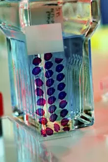
Biopsy slides F007 / 0316
Biopsy slides

Background imageHistopathology Collection: Acute myeloid leukaemia, micrograph

Acute myeloid leukaemia, micrograph
Acute myeloid leukaemia. Light micrograph of blood cells from bone marrow in a case of acute myeloid leukaemia. These cells include a monocytoid myeloblast stem cell

Background imageHistopathology Collection: Elliptocytosis, light micrograph

Elliptocytosis, light micrograph
Elliptocytosis. Light micrograph of red blood cells in a case of elliptocytosis. Red blood cells (erythrocytes) carry oxygen and carbon dioxide to and from body tissues

Background imageHistopathology Collection: Cystine in bone marrow, light micrograph

Cystine in bone marrow, light micrograph
Cystine in bone marrow. Light micrograph of crystals of cystine among blood cells in a sample of bone marrow. Cystine is an amino acid that can form crystals in urine

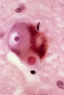
Background imageHistopathology Collection: Parkinsons disease, light micrograph

Parkinsons disease, light micrograph
Parkinsons disease. Light micrograph of a section through the sabstantia nigra of the brain of a patient with Parkinsons disease showing a neuron (nerve cell)

Background imageHistopathology Collection: Dementia, light micrograph

Dementia, light micrograph
Dementia. Light micrograph of a section through the brain of a patient with dementia. Dementia is mainly a disease of old age

Background imageHistopathology Collection: Pernicious anaemia, light micrograph

Pernicious anaemia, light micrograph
Pernicious anaemia. Light micrograph of megaloblast blood cells from bone marrow in a case of pernicious anaemia, also known as Biermers anaemia

Background imageHistopathology Collection: Blood cell cancer, light micrograph

Blood cell cancer, light micrograph
Blood cell cancer. Light micrograph of blood cells from a lymphatic ganglion in a case of a blood cancer of a mixed cell type

Background imageHistopathology Collection: Infectious mononucleosis, micrograph

Infectious mononucleosis, micrograph
Infectious mononucleosis. Light micrograph of blood cells obtained from an inflamed lymphatic ganglion in a case of infectious mononucleosis (glandular fever)

Background imageHistopathology Collection: Lymphoblastic sarcoma, light micrograph

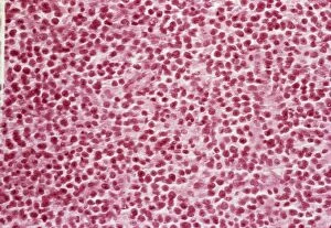
Lymphoblastic sarcoma, light micrograph
Lymphoblastic sarcoma. Light micrograph of blood cells from bone marrow in a case of lymphoblastic sarcoma. These small lymphoblasts (dark red) characteristically have conspicuous nucleoli

Background imageHistopathology Collection: Multiple myeloma, light micrograph

Multiple myeloma, light micrograph
Multiple myeloma. Light micrograph of dystrophy (abnormality) of plasmocyte blood cells from bone marrow in a case of multiple myeloma, also known as Kahlers disease or plasma cell myeloma

Background imageHistopathology Collection: Cushings syndrome, light micrograph

Cushings syndrome, light micrograph
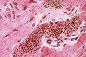
Cushings syndrome. Light micrograph of a section through muscle affected by Cushings syndrome, a hormonal condition caused by overproduction of adrenal gland hormones

Background imageHistopathology Collection: Lymphoplasmacytic lymphoma, micrograph

Lymphoplasmacytic lymphoma, micrograph
Lymphoplasmacytic lymphoma. Light micrograph of a mixture of small lymphocytes and plasma cells from bone marrow in a case of lymphoplasmacytic lymphoma, also known as Waldenstroms macroglobulinemia

Background imageHistopathology Collection: Hairy cell leukaemia, light micrograph

Hairy cell leukaemia, light micrograph
Hairy cell leukaemia. Light micrograph of blood cells in a case of hairy cell leukaemia. This sample, treated with May-Grunwald-Giemsa (MGG) stain

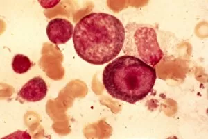
Background imageHistopathology Collection: Acute erythroid leukaemia, micrograph

Acute erythroid leukaemia, micrograph
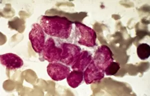
Acute erythroid leukaemia. Light micrograph of blood cells from bone marrow in a case of acute erythroid leukaemia. The cells include dystrophic (degenerated) erythroblasts (nuclei stained dark red)

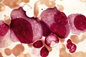
Background imageHistopathology Collection: Acute lymphoblastic leukaemia, micrograph

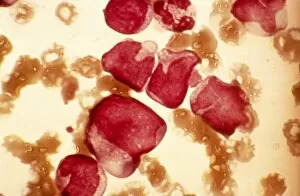
Acute lymphoblastic leukaemia, micrograph
Acute lymphoblastic leukaemia. Light micrograph of blood cells from bone marrow in a case of acute lymphoblastic leukaemia. Leukaemia is a cancer where certain blood cells form in excess

Background imageHistopathology Collection: Malignant histiocytosis, light micrograph

Malignant histiocytosis, light micrograph
Malignant histiocytosis. Light micrograph of blood cells in a case of malignant histiocytosis. Hystiocytosis is an excess number of histiocytes

Background imageHistopathology Collection: Chediak-Higashi syndrome, micrograph

Chediak-Higashi syndrome, micrograph
Chediak-Higashi syndrome. Light micrograph of blood cells in a case of Chediak-Higashi syndrome. The cells include neutrophil white blood cells with abnormal granules

Background imageHistopathology Collection: Metastatic breast cancer, micrograph

Metastatic breast cancer, micrograph
Metastatic breast cancer. Light micrograph of blood cells in a case of metastatic (secondary) medullary breast cancer. Medullary breast cancer is a rare form of breast cancer that is diagnosed by

Background imageHistopathology Collection: Metastatic lung cancer, micrograph

Metastatic lung cancer, micrograph
Metastatic lung cancer. Light micrograph of blood cells in a case of metastatic (secondary) medullary lung cancer. Medullary cancer (carcinoma)

Background imageHistopathology Collection: Multiple sclerosis, light micrograph

Multiple sclerosis, light micrograph
Multiple sclerosis. Light micrograph of a section through the brain showing a large area of demyelinated nerve fibres (light pink, top) due to multiple sclerosis

Background imageHistopathology Collection: Alders anomaly, light micrograph

Alders anomaly, light micrograph
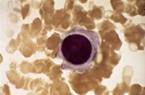
Alders anomaly. Light micrograph of an abnormal lymphocyte white blood cell (centre) from a blood sample in a case of Alders anomaly

Background imageHistopathology Collection: Acute leukaemia, light micrograph

Acute leukaemia, light micrograph
Acute leukaemia. Light micrograph of blood cells from bone marrow in a case of acute leukaemia. These cells include a polynuclear degranulated basophil and myeloblasts

Background imageHistopathology Collection: Chronic myeloid leukaemia, micrograph

Chronic myeloid leukaemia, micrograph
Chronic myeloid leukaemia. Light micrograph of blood cells from bone marrow in a case of chronic myeloid leukaemia. Leukaemia is a cancer where certain blood cells form in excess

Background imageHistopathology Collection: Reticulosarcoma, light micrograph

Reticulosarcoma, light micrograph
Reticulosarcoma. Light micrograph of reticulocyte blood cells (red, one at upper left) from a lymphatic ganglion in a case of reticulosarcoma

Background imageHistopathology Collection: Langerhans cell histiocytosis, micrograph

Langerhans cell histiocytosis, micrograph
Langerhans cell histiocytosis. Light micrograph of blood cells in a case of Langerhans cell histiocytosis (LCH), also called X-type histiocytosis

Background imageHistopathology Collection: Hodgkins disease, light micrograph

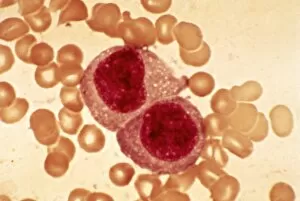
Hodgkins disease, light micrograph
Hodgkins disease. Light micrograph of two non-dystrophic histiocyte blood cells (centre) in a case of Hodgkins disease, also called Hodgkins lymphoma

Background imageHistopathology Collection: Reticulosarcoma, light micrograph C015 / 7133

Reticulosarcoma, light micrograph C015 / 7133
Reticulosarcoma. Light micrograph of reticulocyte blood cells (red, one at upper left) from a lymphatic ganglion in a case of reticulosarcoma

Background imageHistopathology Collection: Brenner tumour of the ovary C015 / 7137

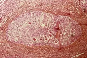
Brenner tumour of the ovary C015 / 7137
Brenner tumour of the ovary. Light micrograph of a section through an ovary showing a Brenner tumour (centre). This rare benign (non-cancerous)

Background imageHistopathology Collection: Langerhans cell histiocytosis C015 / 7135

Langerhans cell histiocytosis C015 / 7135
Langerhans cell histiocytosis. Light micrograph of a section through a lymph node showing Langerhans cell histiocytosis (LCH), also known as granulomatosis

Background imageHistopathology Collection: Sarcoidosis, light micrograph C015 / 7134

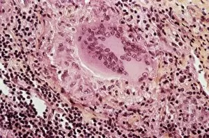
Sarcoidosis, light micrograph C015 / 7134
Sarcoidosis. Light micrograph of a section through a lymph node affected by sarcoidosis. Also known as Boecks disease, this is a chronic inflammation of the lymph nodes or organs

Background imageHistopathology Collection: Lung silicosis, light micrograph C015 / 7129

Lung silicosis, light micrograph C015 / 7129
Lung silicosis. Light micrograph of a section through a lung with silicosis, a type of pneumoconiosis caused by exposure to silica dusts. The lung tissue has become fibrous (centre)

Background imageHistopathology Collection: Thymus gland cancer, light micrograph C015 / 7128

Thymus gland cancer, light micrograph C015 / 7128
Thymus cancer. Light micrograph of a section through a thymus gland with thymoma cancer. The thymus is a gland in the upper chest that plays an important role in the immune system

Background imageHistopathology Collection: Endometrial hyperplasia, light micrograph C015 / 7104

Endometrial hyperplasia, light micrograph C015 / 7104
Endometrial hyperplasia. Light micrograph of a section through tissue from the endometrium, in a case of endometrial hyperplasia. The endometrium is the uterus lining


All Professionally Made to Order for Quick Shipping

Histopathology is a fascinating field that delves into the intricate details of diseases and their impact on our bodies. From Salmonella bacteria to Dohle bodies in blood cells, histopathologists use various techniques like SEM (scanning electron microscopy) and micrographs to unravel the mysteries within. In acute promyelocytic leukemia, abnormal cells can be observed through micrographs, providing crucial insights for diagnosis and treatment. Similarly, ovarian cancer reveals its telltale signs under light micrograph C015 / 7103, aiding in early detection. Liver tissue cirrhosis showcases the damaging effects of certain conditions on this vital organ. Through light micrographs, pathologists can identify key features that aid in understanding disease progression. Cystic fibrosis affects multiple organs including the lungs and pancreas. Histopathological analysis helps uncover cellular changes associated with this genetic disorder. Osteoporotic bone exhibits reduced density and weakened structure due to bone loss over time. By studying these samples closely, researchers gain valuable knowledge about prevention strategies and potential treatments. Malaria parasites are intricately studied using TEM (transmission electron microscopy), revealing their distinct characteristics necessary for accurate diagnosis and effective interventions against this deadly disease. Herpes simplex viruses leave their mark through TEM images showcasing their unique structures. Understanding these viral particles aids in developing antiviral therapies to combat outbreaks effectively. Diabetic foot ulcers present complex challenges due to impaired wound healing mechanisms caused by diabetes-related complications. Histopathology plays a pivotal role in unraveling the underlying pathophysiology behind these ulcers for better management strategies. Gout crystals provide visual evidence of joint inflammation caused by excess uric acid deposition. Studying these crystals enables clinicians to tailor treatment plans accordingly for patients suffering from gout attacks. HIV particles reveal themselves under TEM examination as we strive towards understanding this global health crisis better. This microscopic exploration contributes significantly towards developing effective antiretroviral therapies and potential vaccines.