
Human Biology Collection
"Exploring the Wonders of Human Biology: From Normal Knees to Nerve Cells" Unveiling the intricate beauty of human biology


All Professionally Made to Order for Quick Shipping
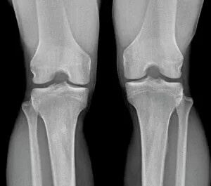
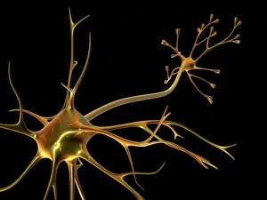
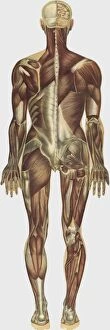
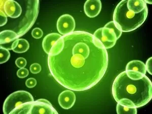
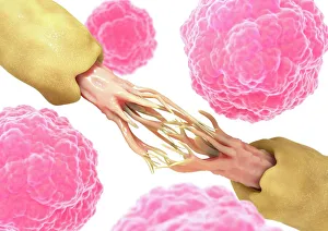
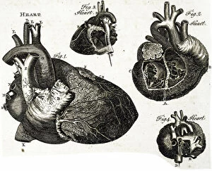
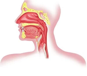
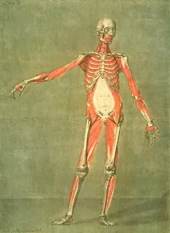
"Exploring the Wonders of Human Biology: From Normal Knees to Nerve Cells" Unveiling the intricate beauty of human biology, starting with a glimpse into our normal knees through X-ray imaging. Delving deeper into our most complex organ, the brain, as MRI scans reveal its fascinating structure and functionality in all its normal glory. Conceptual artwork depicting knee pain serves as a reminder of the challenges we face within our own bodies and highlights the importance of understanding their mechanisms. Witnessing the awe-inspiring complexity of nerve cells through stunning artwork that captures their intricate networks and vital role in transmitting information throughout our body. Exploring running injuries through conceptual artwork, shedding light on common ailments faced by athletes and emphasizing the need for proper care and prevention techniques. Admiring an artistic representation of the human heart, symbolizing life's powerful engine that beats tirelessly to sustain us every day. Julien Bougle's masterpiece showcases colored plates superimposed on a human body, offering a unique perspective on how different systems intertwine harmoniously within us. Peering into an X-ray image revealing a normal child's head reminds us of nature's remarkable design while instilling hope for healthy futures ahead. Conceptual computer artwork brings cells to life before our eyes, unveiling their microscopic world filled with astonishing diversity and essential functions crucial for survival. Examining leptin - a molecular model representing this hormone responsible for regulating appetite - unveils yet another piece in humanity's biological puzzle towards better understanding nutrition-related disorders like obesity or anorexia nervosa. Artwork portraying nerve damage alongside stem cells sparks curiosity about potential therapeutic approaches harnessing these regenerative powerhouses to repair injured nerves and restore function effectively. A mesmerizing portrayal of nerve cells invites contemplation about their immense significance in orchestrating communication between various bodily systems – truly the conductors of our biological symphony.