
Immunity Collection
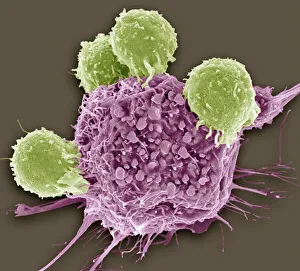
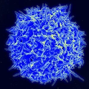
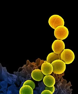
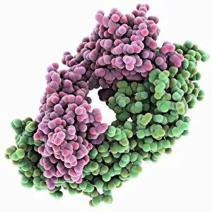
"Unlocking the Power of Immunity: A Journey into Cellular Defense" In the intricate world of immunity, T lymphocytes emerge as fearless warriors against cancer cells


All Professionally Made to Order for Quick Shipping
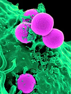
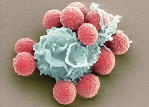
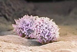
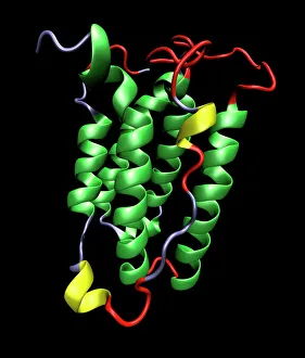
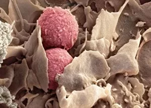
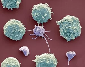
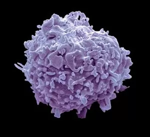
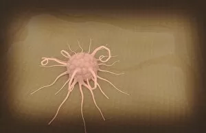
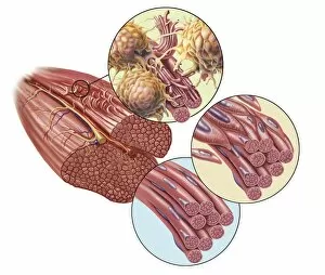
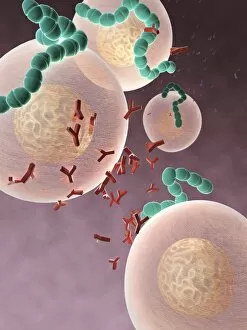
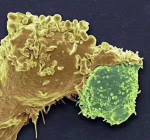
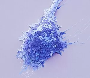
"Unlocking the Power of Immunity: A Journey into Cellular Defense" In the intricate world of immunity, T lymphocytes emerge as fearless warriors against cancer cells. SEM C001 / 1679 reveals their relentless pursuit in eradicating these malignant invaders. Neutrophils, our unsung heroes, engulf MRSA bacteria with unwavering determination. Witness this extraordinary battle through the lens of SEM C018 / 8596, where a neutrophil devours its prey. Blood cells flow through our veins like an army on standby, ready to defend us at any moment. Their diverse roles and functions make them indispensable guardians of our health. Plasma cells take center stage under TEM's gaze, showcasing their remarkable ability to produce antibodies that neutralize threats lurking within us. Phagocytosis becomes an art form when fungal spores fall victim to its grasp. Observe this captivating spectacle captured by SEM's lens—a testament to nature's resilience. Macrophages stand tall as they engulf TB bacteria in a fierce struggle for survival. Through SEM imagery and sheer determination, we witness their valiant efforts to protect us from harm. The interferon molecule emerges as a beacon of hope amidst viral chaos—an essential messenger that rallies immune responses against invading pathogens. Activated macrophages proudly display their formidable armor under SEM C015 / 6375—proof of their readiness to combat any intruders threatening our well-being. TEM unveils the inner workings of macrophage cells—their intricate machinery designed for surveillance and destruction—a constant reminder that vigilance is key in maintaining immunity. Once again, we witness macrophages engaging in battle against TB bacteria through the lens of SEM—an awe-inspiring sight that reaffirms the power within each cell fighting for our defense. Amidst frozen Baltic waters, a man immerses himself—a symbolic act echoing nature's ability to invigorate and strengthen both body and mind—a reminder of the resilience we possess.