
Immunofluores Collection
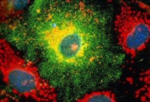
"Unveiling the Intricacies of Cells: A Journey through Immunofluorescence" Immunofluorescent LM, a powerful technique


All Professionally Made to Order for Quick Shipping
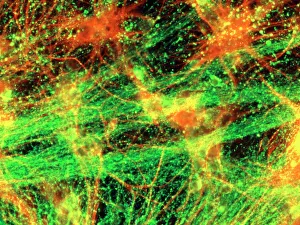
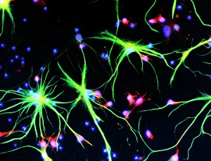
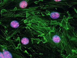
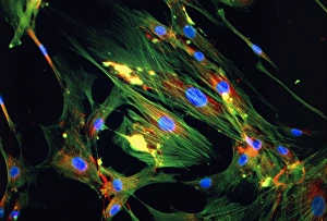
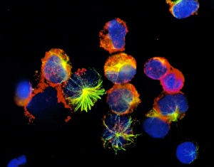
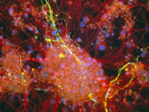
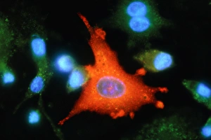
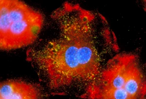
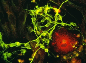
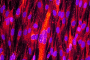
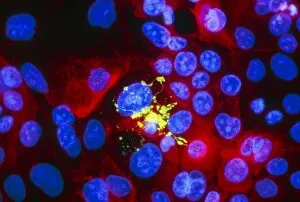
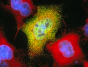
"Unveiling the Intricacies of Cells: A Journey through Immunofluorescence" Immunofluorescent LM, a powerful technique, allows us to delve into the mesmerizing world of cells. With its aid, we can visualize and understand their complex structures and functions. Nerve cells, also known as neurons, light up under immunofluorescent LM revealing their intricate networks. These vital components of our nervous system play a crucial role in transmitting information throughout our body. Astrocytes, another type of cell found in the brain and spinal cord, are brought to life through immunofluorescence. Their star-like shape becomes apparent as they support and nourish neurons. Moving beyond nerve cells, fibroblast cell nuclei come into focus under immunofluorescent LM. These versatile cells are responsible for producing connective tissue proteins essential for wound healing and maintaining skin elasticity. Skin fibroblast cells take center stage next as immunofluorescence highlights their presence within our largest organ. Understanding these cells aids in unraveling the mysteries behind various skin conditions and diseases. Immunofluorescent LM unveils normal breast cells with precision. This knowledge is invaluable when studying breast cancer development or identifying potential therapeutic targets. Natural killer blood cells shine brightly under immunofluorescence microscopy. Exploring these immune warriors provides insights into how they detect and eliminate infected or cancerous cells within our body. Keratinocyte skin cells reveal their beauty through immunofluorescent LM techniques. By understanding these building blocks of our epidermis, researchers gain valuable knowledge about skin disorders such as psoriasis or eczema. The wonders continue with rat brain cells and axons illuminated by immunofluorescence microscopy. Studying these structures helps uncover mechanisms underlying neurological disorders like Alzheimer's disease or Parkinson's disease. An active macrophage takes center stage under the fluorescent glow bestowed by immunofluorescent LM.