Fraud Blocker Skip to main content

Immunology Collection

"Unleashing the Power of Immunology: Exploring the Intricate World of Immune Responses" Immunology

Background imageImmunology Collection: Immunoglobulin G antibody molecule

Immunoglobulin G antibody molecule. Computer model of the secondary structure of immunoglobulin G (IgG). This is the most abundant immunoglobulin and is found in all body fluids

Background imageImmunology Collection: T lymphocytes and cancer cell, SEM C001 / 1679

T lymphocytes and cancer cell, SEM C001 / 1679
T lymphocytes and cancer cell. Coloured scanning electron micrograph (SEM) of T lymphocyte cells (green) attached to a cancer cell

Background imageImmunology Collection: Immunoglobulin G antibody molecule F007 / 9894

Immunoglobulin G antibody molecule F007 / 9894
Immunoglobulin G antibody molecule. Computer model of the secondary structure of immunoglobulin G (IgG). This is the most abundant immunoglobulin and is found in all body fluids

Background imageImmunology Collection: Neutrophil engulfing MRSA, SEM C018 / 8596

Neutrophil engulfing MRSA, SEM C018 / 8596
Neutrophil engulfing MRSA. Coloured scanning electron micrograph (SEM) of a neutrophil white blood cell (green) engulfing methicillin-resistant Staphylococcus aureus bacteria (MRSA, pink)

Background imageImmunology Collection: Dendritic cells, artwork

Dendritic cells, artwork. Dendritic cells, a type of white blood cell, are called antigen presenting cells (APCs) and are found in the bodys tissues

Background imageImmunology Collection: TEM of human white blood cell bearing HLA antigen

TEM of human white blood cell bearing HLA antigen
False-colour transmission electron micrograph (TEM) of a human white blood cell (bottom), showing presence of an HLA antigen (human leucocyte antigen)

Background imageImmunology Collection: Antibodies, artwork

Antibodies, artwork
Computer artwork of antibody molecules showing the structure of an immunoglobulin G (IgG) molecule. This is the most abundant immunoglobulin and is found in all body fluids

Background imageImmunology Collection: Bacteria infecting a macrophage, SEM

Bacteria infecting a macrophage, SEM
Bacteria infecting a macrophage. Coloured scanning electron micrograph (SEM) of Mycobacterium tuberculosis bacteria (purple) infecting a macrophage white blood cell

Background imageImmunology Collection: HIV reverse transcription enzyme

HIV reverse transcription enzyme. Molecular models of the reverse transcriptase enzyme found in HIV (the human immunodeficiency virus)

Background imageImmunology Collection: TEM of human macrophage

TEM of human macrophage

Background imageImmunology Collection: Basophil white blood cell

Basophil white blood cell. Coloured transmission electron micrograph (TEM) of a basophil white blood cell. Basophils are the smallest and rarest of the white blood cells

Background imageImmunology Collection: Dohle bodies in blood cell, micrograph

Dohle bodies in blood cell, micrograph
Dohle bodies in blood cell. Light micrograph of a neutrophil white blood cell (centre) with Dohle bodies. These are small inclusions within the cells cytoplasm

Background imageImmunology Collection: Macrophage engulfing TB bacteria, SEM

Macrophage engulfing TB bacteria, SEM
Macrophage engulfing TB bacteria. Coloured scanning electron micrograph (SEM) of a macrophage white blood cell (purple) engulfing a tuberculosis (Mycobacterium tuberculosis) bacterium (pink)

Background imageImmunology Collection: Activated macrophage, SEM C015 / 6375

Activated macrophage, SEM C015 / 6375
Activated macrophage. Coloured scanning electron micrograph (SEM) of an active macrophage white blood cell. Macrophages are cells of the bodys immune system that are found in the tissues rather than

Background imageImmunology Collection: Plasma cells, TEM

Plasma cells, TEM
Plasma cells. Coloured transmission electron micrograph (TEM) of plasma cells. Plasma cells are mature B lymphocytes (white blood cells) that produce and secrete antibodies during an immune response

Background imageImmunology Collection: Acute promyelocytic leukaemia, micrograph

Acute promyelocytic leukaemia, micrograph
Acute promyelocytic leukaemia. Light micrograph of blood cells from bone marrow in a case of acute promyelocytic leukaemia. Leukaemia is a cancer where certain blood cells form in excess

Background imageImmunology Collection: Phagocytosis of fungal spores, SEM

Phagocytosis of fungal spores, SEM
Phagocytosis of a fungus spore. Coloured scanning electron micrograph (SEM) of a neutrophil (blue) phagocytosing (engulfing and destroying) spores from the fungus Aspergillus fumigatus (red)

Background imageImmunology Collection: Neutrophil cell trapping bacteria, SEM

Neutrophil cell trapping bacteria, SEM
Neutrophil cell trapping bacteria. Coloured scanning electron micrograph (SEM) of bacteria (rod-shaped) being trapped by a neutrophil cell

Background imageImmunology Collection: Macrophage cells, SEM

Macrophage cells, SEM
Macrophage cells. Coloured scanning electron micrograph (SEM) of macrophage white blood cells. Macrophages are cells of the bodys immune system

Background imageImmunology Collection: Interferon molecule

Interferon molecule. Computer model showing the secondary structure of a molecule of interferon. Interferons are proteins produced by white blood cells as part of the immune response to invading

Background imageImmunology Collection: Macrophage engulfing bead

Macrophage engulfing bead
Illustration based on a scanning electron micrograph of a human macrophage phagocytosing (engulfing) a latex bead. Macrophages are monocytes from circulating blood that have migrated to tissues

Background imageImmunology Collection: Lymphocyte white blood cells, artwork

Lymphocyte white blood cells, artwork
Lymphocyte white blood cells. Computer artwork of lymphocyte white blood cells, showing their nuclei (red, centre). Lymphocytes, like all white blood cells, are part of the immune system

Background imageImmunology Collection: Bacteria infecting a macrophage, SEM

Bacteria infecting a macrophage, SEM
Bacteria infecting a macrophage. Coloured scanning electron micrograph (SEM) of Mycobacterium tuberculosis bacteria (purple) infecting a macrophage white blood cell

Background imageImmunology Collection: Female body showing digestive and circulatory system

Female body showing digestive and circulatory system
Medical illustration of female body showing digestive and circulatory system, perspective view

Background imageImmunology Collection: Medicine. Edward Jenner and the smallpox vaccine. Imagery, France, c.1900

Medicine. Edward Jenner and the smallpox vaccine. Imagery, France, c.1900
2636415 Medicine. Edward Jenner and the smallpox vaccine. Imagery, France, c.1900.; Private Collection; (add.info.: Medecine. Coll. Part.); Photo © CCI.

Background imageImmunology Collection: Paul Ehrlich

Paul Ehrlich
3253013 Paul Ehrlich; (add.info.: Paul Ehrlich , 1854 -1915. German physician and scientist. From Enciclopedia Ilustrada Segui, published c. 1900).

Background imageImmunology Collection: Louis Pasteur (1822-1895) French chemist, in his laboratory at the Ecole Normale, Paris

Louis Pasteur (1822-1895) French chemist, in his laboratory at the Ecole Normale, Paris
527302 Louis Pasteur (1822-1895) French chemist, in his laboratory at the Ecole Normale, Paris, during his work on hydrophobia

Background imageImmunology Collection: Army, U.S. Army Medical School; Typhoid Vaccine, 1917. Creator: Harris & Ewing. Army, U.S

Army, U.S. Army Medical School; Typhoid Vaccine, 1917. Creator: Harris & Ewing. Army, U.S
Army, U.S. Army Medical School; Typhoid Vaccine, 1917

Background imageImmunology Collection: Army, U.S. Army Medical School; Typhoid Vaccine, 1917. Creator: Harris & Ewing. Army, U.S

Army, U.S. Army Medical School; Typhoid Vaccine, 1917. Creator: Harris & Ewing. Army, U.S
Army, U.S. Army Medical School; Typhoid Vaccine, 1917

Background imageImmunology Collection: Army, U.S. Army Medical School; Typhoid Vaccine, 1917. Creator: Harris & Ewing. Army, U.S

Army, U.S. Army Medical School; Typhoid Vaccine, 1917. Creator: Harris & Ewing. Army, U.S
Army, U.S. Army Medical School; Typhoid Vaccine, 1917

Background imageImmunology Collection: Army, U.S. Army Medical School; Typhoid Vaccine, 1917. Creator: Harris & Ewing. Army, U.S

Army, U.S. Army Medical School; Typhoid Vaccine, 1917. Creator: Harris & Ewing. Army, U.S
Army, U.S. Army Medical School; Typhoid Vaccine, 1917

Background imageImmunology Collection: Army, U.S. Army Medical School; Typhoid Vaccine, 1917. Creator: Harris & Ewing. Army, U.S

Army, U.S. Army Medical School; Typhoid Vaccine, 1917. Creator: Harris & Ewing. Army, U.S
Army, U.S. Army Medical School; Typhoid Vaccine, 1917

Background imageImmunology Collection: Army, U.S. Army Medical School; Typhoid Vaccine, 1917. Creator: Harris & Ewing. Army, U.S

Army, U.S. Army Medical School; Typhoid Vaccine, 1917. Creator: Harris & Ewing. Army, U.S
Army, U.S. Army Medical School; Typhoid Vaccine, 1917

Background imageImmunology Collection: Army, U.S. Army Medical School; Typhoid Vaccine, 1917. Creator: Harris & Ewing. Army, U.S

Army, U.S. Army Medical School; Typhoid Vaccine, 1917. Creator: Harris & Ewing. Army, U.S
Army, U.S. Army Medical School; Typhoid Vaccine, 1917

Background imageImmunology Collection: Army, U.S. Army Medical School; Typhoid Vaccine, 1917. Creator: Harris & Ewing. Army, U.S

Army, U.S. Army Medical School; Typhoid Vaccine, 1917. Creator: Harris & Ewing. Army, U.S
Army, U.S. Army Medical School; Typhoid Vaccine, 1917

Background imageImmunology Collection: Army, U.S. Army Medical School; Typhoid Vaccine, 1917. Creator: Harris & Ewing. Army, U.S

Army, U.S. Army Medical School; Typhoid Vaccine, 1917. Creator: Harris & Ewing. Army, U.S
Army, U.S. Army Medical School; Typhoid Vaccine, 1917

Background imageImmunology Collection: Japanese relocation, California, 1942. Creator: Dorothea Lange

Japanese relocation, California, 1942. Creator: Dorothea Lange
Japanese relocation, California. Newcomers at Santa Anita Park Assembly Center in Arcadia, California, receive vaccination against smallpox and typhoid fever. Under direction of Dr

Background imageImmunology Collection: Paul Ehrlich, German doctor and scientist (b / w photo)

Paul Ehrlich, German doctor and scientist (b / w photo)
6014957 Paul Ehrlich, German doctor and scientist (b/w photo) by Perscheid, Nicola (1864-1930); Private Collection; (add.info.: Paul Ehrlich (1854-1915)

Background imageImmunology Collection: Rev. Cotton Mather (Son of Increase) Using His Powerful Influence to Overcome

Rev. Cotton Mather (Son of Increase) Using His Powerful Influence to Overcome the Prejudice Against Inoculation for Smallpox in Boston, 1721, before 1926

Background imageImmunology Collection: Paul Ehrlich, German doctor and scientist (b / w photo)

Paul Ehrlich, German doctor and scientist (b / w photo)
6014954 Paul Ehrlich, German doctor and scientist (b/w photo) by German Photographer (20th Century); Private Collection; (add.info.: Paul Ehrlich (1854-1915)

Background imageImmunology Collection: Edward Jenner, English doctor and discoverer of a vaccine for smallpox. (chromolitho)

Edward Jenner, English doctor and discoverer of a vaccine for smallpox. (chromolitho)
668844 Edward Jenner, English doctor and discoverer of a vaccine for smallpox. (chromolitho) by French School, (19th century); Private Collection; (add.info.: Edward Jenner)

Background imageImmunology Collection: Louis Pasteur and his collaborators in working on a vaccine for croup at the Institut Pasteur in

Louis Pasteur and his collaborators in working on a vaccine for croup at the Institut Pasteur in Paris (b / w photo)
3098643 Louis Pasteur and his collaborators in working on a vaccine for croup at the Institut Pasteur in Paris (b/w photo) by French Photographer

Background imageImmunology Collection: Peaceful glories of France (colour litho)

Peaceful glories of France (colour litho)
3099324 Peaceful glories of France (colour litho) by French School, (19th century); Private Collection; (add.info.: Peaceful glories of France)

Background imageImmunology Collection: Jean-Baptiste Jupille, French shepherd boy who fought off a rabid dog to prevent it attacking

Jean-Baptiste Jupille, French shepherd boy who fought off a rabid dog to prevent it attacking other children
974398 Jean-Baptiste Jupille, French shepherd boy who fought off a rabid dog to prevent it attacking other children (chromolitho) by European School

Background imageImmunology Collection: Emil von Behring, German doctor and physiologist, c1890 (b / w photo)

Emil von Behring, German doctor and physiologist, c1890 (b / w photo)
5232155 Emil von Behring, German doctor and physiologist, c1890 (b/w photo) by German Photographer, (19th century); Private Collection; (add.info.: Emil von Behring (1854-1917)

Background imageImmunology Collection: Edward Jenner (chromolitho)

Edward Jenner (chromolitho)
961327 Edward Jenner (chromolitho) by English School, (19th century); Private Collection; (add.info.: Edward Jenner, born in 1749, discovered the vaccine in 1796, died in 1823)

Background imageImmunology Collection: Edward Jenner, English physician and scientist (engraving)

Edward Jenner, English physician and scientist (engraving)
979428 Edward Jenner, English physician and scientist (engraving) by Marshall, William Calder (1813-94); Private Collection; (add.info.: Edward Jenner (1749-1823), English physician and scientist

Background imageImmunology Collection: Elie Metchnikoff, Russian zoologist (colour litho)

Elie Metchnikoff, Russian zoologist (colour litho)
3620555 Elie Metchnikoff, Russian zoologist (colour litho) by Barrere, Adrien (1877-1931); Private Collection; (add.info.: Elie Metchnikoff (1845-1916), Russian zoologist.); © Look and Learn


All Professionally Made to Order for Quick Shipping

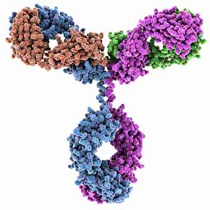
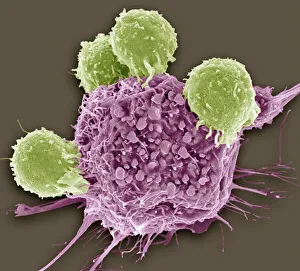
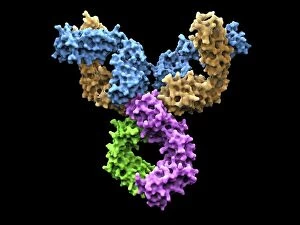
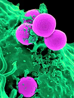
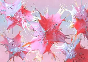
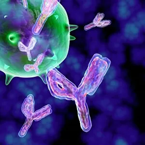
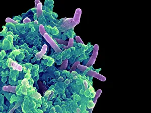
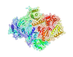
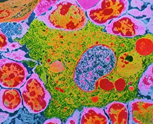
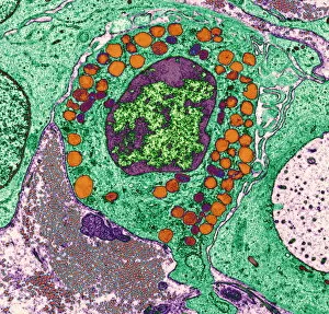
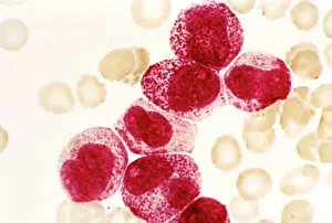
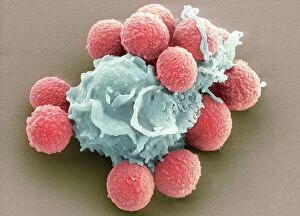
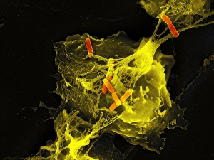
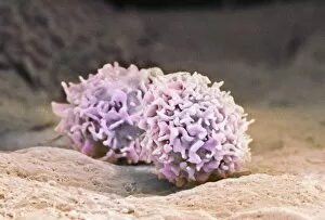
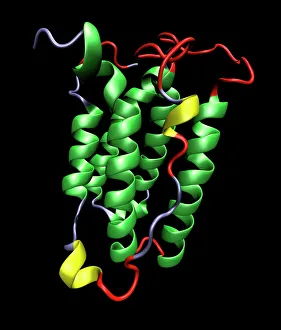
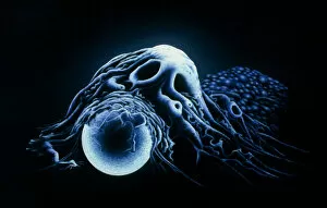
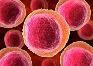
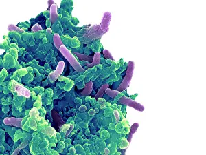
"Unleashing the Power of Immunology: Exploring the Intricate World of Immune Responses" Immunology, a fascinating field that delves into the complex mechanisms of our immune system, holds immense potential in combating diseases. T lymphocytes and cancer cells engage in a constant battle for supremacy, as depicted by SEM C001 / 1679. These tiny warriors play a crucial role in identifying and eliminating abnormal cells. The Immunoglobulin G antibody molecule (F007 / 9894) stands tall as one of our body's most powerful defenders against pathogens. Its remarkable structure enables it to neutralize harmful invaders with precision and efficiency. Neutrophils, exemplified by SEM C018 / 8596, showcase their extraordinary ability to engulf MRSA bacteria—an awe-inspiring sight indeed. Meanwhile, dendritic cells (artwork) act as vigilant sentinels, capturing antigens and presenting them to other immune cells for recognition. TEM reveals an up-close view of human white blood cells bearing HLA antigens—a key component in distinguishing self from non-self. Antibodies (artwork), resembling elegant warriors on a mission, bind specifically to foreign substances to mark them for destruction. HIV reverse transcription enzyme serves as a reminder of the challenges faced by immunologists worldwide. This relentless virus exploits our own cellular machinery but continues to be targeted through innovative research efforts. Human macrophages (TEM) demonstrate their exceptional phagocytic abilities while basophil white blood cells stand ready at the frontlines—both integral players in mounting effective immune responses against invading pathogens. Intriguingly captured by SEM imagery is bacteria infecting a macrophage—a visual representation highlighting how these microscopic organisms can exploit host defenses while also serving as valuable tools for studying infection dynamics. Lastly, Dohle bodies within blood cells offer insights into various pathological conditions affecting neutrophils—an essential clue guiding immunologists towards understanding and treating immune disorders.