
Infections Collection
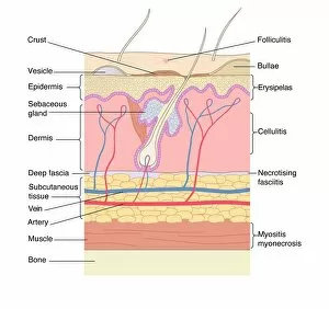
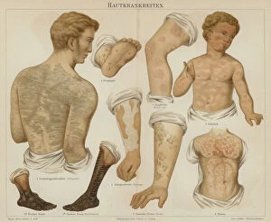
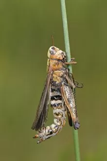
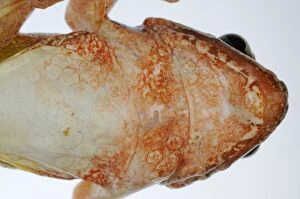
"Infections: Unveiling the Intricate World of Microscopic Battles" Skin disorders, artwork: Delicate canvases marred by unseen adversaries


All Professionally Made to Order for Quick Shipping
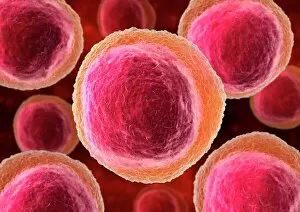
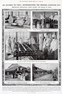
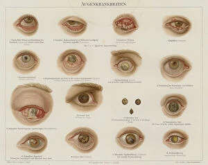
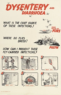
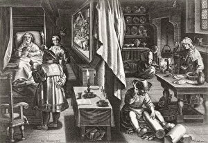
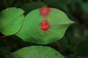
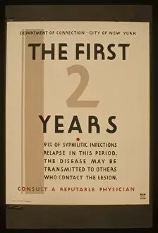
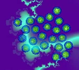
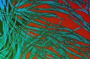
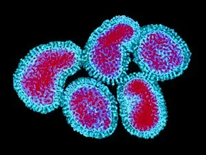
"Infections: Unveiling the Intricate World of Microscopic Battles" Skin disorders, artwork: Delicate canvases marred by unseen adversaries. Rift Valley fever virus, TEM: A glimpse into the minuscule realm where chaos thrives. Lymphocyte white blood cells, artwork: The valiant defenders standing strong against invading hordes. Chlamydia trachomatis bacteria, TEM: Tiny invaders wreaking havoc in unsuspecting hosts. Adenovirus, TEM: Nature's invisible architects of discomfort and distress. Unpredictable future. . Where will the next microbial threat emerge from? Violet bramble rust causing red coloration on Bramble leaf: Nature's artistic touch concealing a hidden menace. WW2 Poster -- Dysentery and Diarrhea: Reminders of past battles fought against insidious foes within our own bodies. Diseases of the throat (colour litho): Silent tormentors lurking in every breath we take. Precautions taken against the plague in Japan, 1908: Historical measures to combat an ancient adversary that still haunts us today. Engraving depicting a doctor examining a baby with measles: Witnessing innocence succumb to microscopic warfare breaks hearts worldwide. Eye disorders (colour litho): Windows to our souls vulnerable to unseen infiltrators. Infections may be invisible but their impact is undeniable – reminding us that even amidst beauty and artistry lies an ongoing battle for survival within our very cells.