mail_outline sales@mediastorehouse.com
Framed Print
Canvas Print
Metal Print
Photographic Print
Poster Print
Fine Art Print
Jigsaw Puzzle
Photo Mug
Greetings Card
Cushion
Mouse Mat
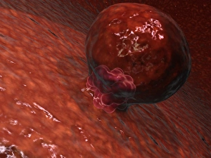
A blastocyst begins implanting in the wall of the uterus
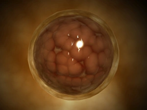
Microscopic view of a blastula during pregnancy. After the cleavage has produced over 100 cells, the embryo is called a blastula
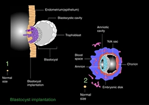
Blastocyst implantation, diagramBlastocyst implantation. Diagram of the blastocyst (lower right), a cellular stage that occurs during human (and all mammalian) reproduction, five days after fertilisation