
Internal Structure Collection
"Exploring the Intricate World Within: Unveiling the Hidden Marvels of Internal Structure" Delving into Nature's Blueprint


All Professionally Made to Order for Quick Shipping
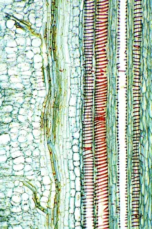
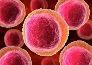
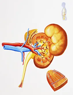
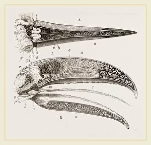
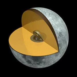
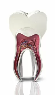
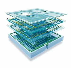
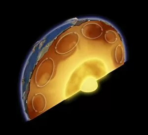
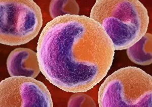
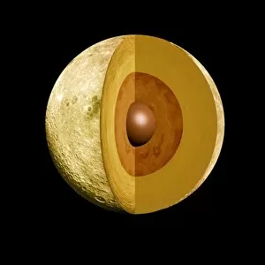
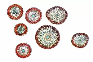
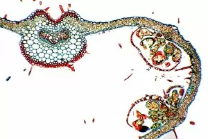
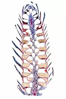
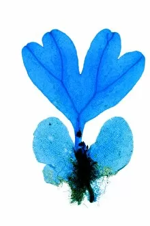
"Exploring the Intricate World Within: Unveiling the Hidden Marvels of Internal Structure" Delving into Nature's Blueprint: A close-up view of a dicotyledon plant stem under a light micrograph reveals its intricate internal structure. The Secret Life of Chloroplasts: An artistic depiction showcases the mesmerizing and complex structure within these vital organelles responsible for photosynthesis. Guardians of Immunity: Lymphocyte white blood cells, portrayed in artwork, exhibit their unique internal structure that aids in defending our bodies against infections. Brewing Perfection: Through scanning electron microscopy (SEM), we uncover the fascinating internal composition of a roasted coffee bean, unveiling its rich flavors and aromas. Unraveling Wood's Mysteries: Colored engravings offer us glimpses into sections of wood's internal structure, showcasing its strength and beauty as nature's building material. Tracing Time through Walnut Trees: Peering into a walnut tree trunk exposes its hidden rings and intricate internal structures, telling tales of growth and resilience over years gone by. Filtering Life's Essence: Examining the internal structure of human kidneys with an inset revealing the medulla highlights their remarkable ability to purify our blood and maintain balance within our bodies. Toucan's Architectural Marvels: Discovering the captivating intricacies within the beak and head of a toucan unveils how evolution has shaped this bird’s unique features for survival in its habitat. Mapping Our Cognitive Centerpiece: A basic cutaway illustration provides insights into the human brain’s inner workings, highlighting key regions like thalamus, cerebrum, and hypothalamus that orchestrate our thoughts and actions. Lunar Landscapes Revealed: Artwork depicting moon structures takes us on an imaginary journey across celestial landscapes while pondering about extraterrestrial mysteries yet to be unraveled. The Framework of Life.