
Intestine Collection
"The Intricate World of Intestines: Exploring the Marvels Within" Delving into the depths of our gastrointestinal system


All Professionally Made to Order for Quick Shipping
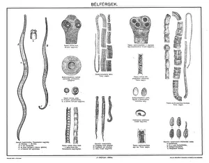
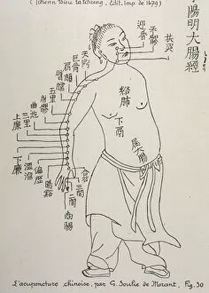
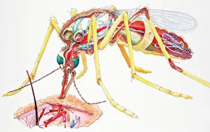
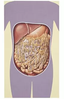
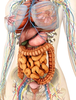
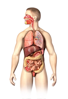
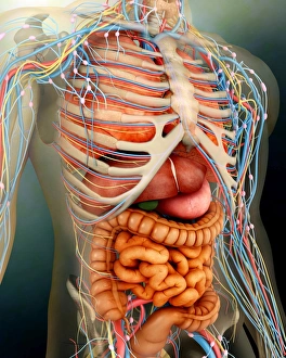
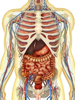
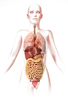
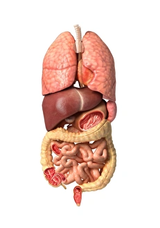
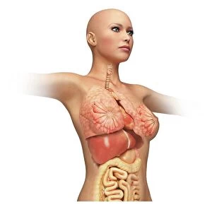
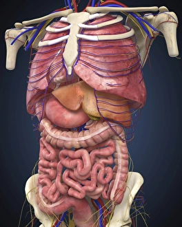
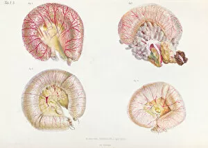
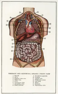
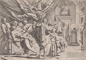
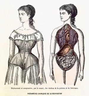
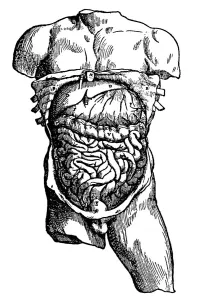
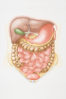
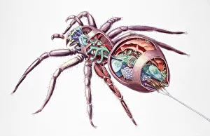
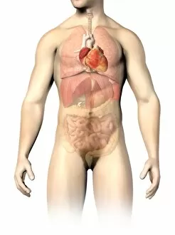
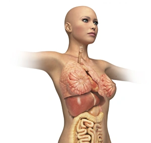
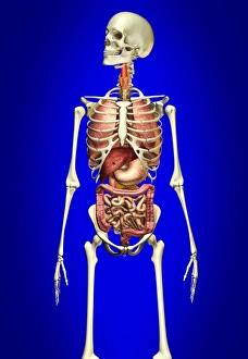
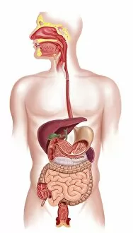
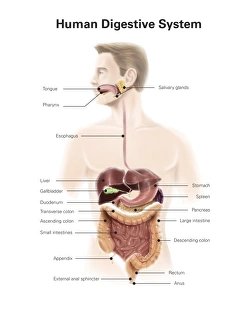
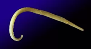
"The Intricate World of Intestines: Exploring the Marvels Within" Delving into the depths of our gastrointestinal system, we uncover a world teeming with life - intestinal parasites known as gastrointestinal nematodes. Did you know that acupuncture can have a positive impact on our intestines? This ancient practice stimulates specific points to promote digestive health and balance. A female mosquito (Culicidae) is not just an annoying pest; its internal anatomy reveals fascinating adaptations for sucking blood from our skin, as seen in a cross-section illustration. The greater omentum, or epiploon, acts as a protective apron within our abdomen. An intriguing historical model showcases its intricate folds and functions. Intestinal protozoan parasites are microscopic organisms that wreak havoc on our gut health. Transmission electron microscopy (TEM) captures their detailed structure and complexity. Parasitic nematode worms may sound like creatures from a horror movie, but they inhabit the intestines of various animals including humans. Understanding their lifecycle is crucial for prevention and treatment. Cryptosporidium protozoa are notorious culprits behind diarrheal diseases worldwide. TEM images provide insights into their unique features and strategies for survival within the intestine. Tiny but mighty - intestinal microvilli play a vital role in nutrient absorption by increasing surface area in the small intestine's lining, as revealed through TEM imaging. Explore an artistic representation of dog anatomy where the intricacies of their intestines come to life through stunning artwork capturing both form and function. The Lazio Roma Rome Velletri Museo Civico di Velletri houses captivating exhibits showcasing ancient Roman medical knowledge related to intestinal health – truly bridging past with present discoveries.