
Ions Collection
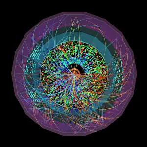
"Ions: Unveiling the Hidden World of Charged Particles" Lead ion collisions: Exploring the mysteries of high-energy particle interactions


All Professionally Made to Order for Quick Shipping
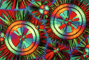
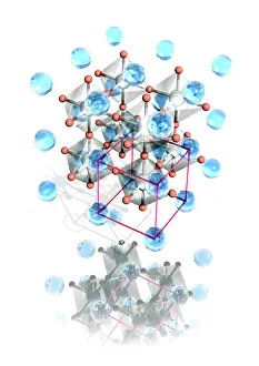
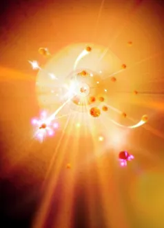
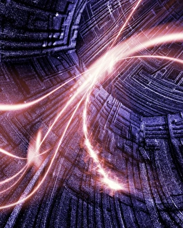
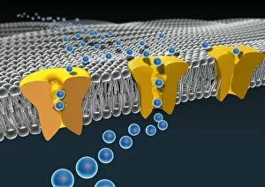
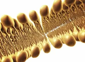
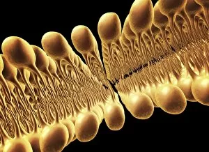
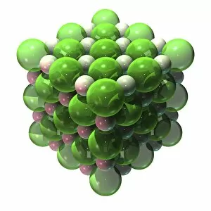
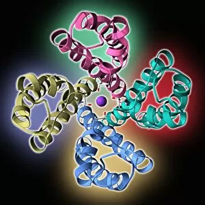
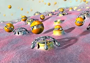
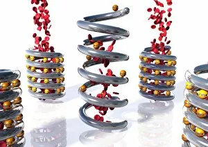
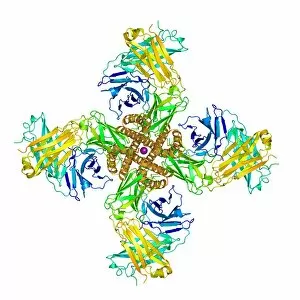
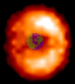
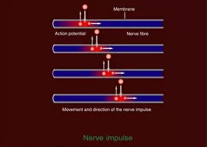
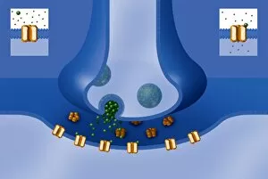
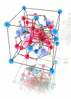
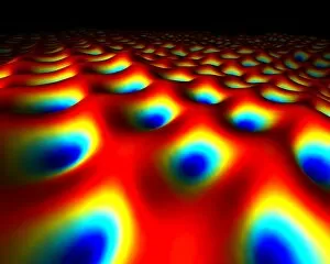
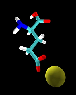
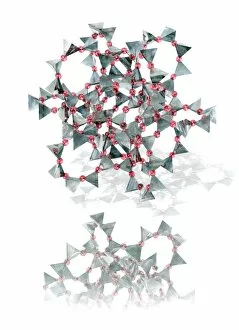
"Ions: Unveiling the Hidden World of Charged Particles" Lead ion collisions: Exploring the mysteries of high-energy particle interactions. Flame tests reveal the vibrant colors produced by different ions in a flame. Magnificent EDTA crystals under a light micrograph, showcasing their intricate beauty. Delving into the fascinating perovskite crystal structure and its unique properties. Witnessing the explosive power of lead ion collisions in cutting-edge particle accelerators. The delicate dance of particles colliding, unraveling secrets at microscopic scales. Journey through time and space with particle accelerators as they unlock new frontiers in scientific discovery. Artistic representation of cell membrane lipid bilayer, highlighting its crucial role in cellular function (artwork F007 / 1477). Revealing the complex network of cell membrane ion channels that regulate vital processes (artwork C016 / 7689). An illustration depicting copper electrode's transformative journey as it reduces hydrogen ions to form hydrogen gas molecules via an external circuit. Close-up view of ball and chain shackles symbolizing freedom from limitations on Oro Grande, California's Route 66 - just like how ions break free from their atomic constraints to shape our world. In this captivating caption, we embark on a scientific odyssey exploring various aspects related to ions – from lead ion collisions and flame tests to EDTA crystals and perovskite structures – unveiling their hidden wonders along with intriguing visuals representing cell membranes, electrodes, and even symbolic imagery evoking liberation itself.