Fraud Blocker Skip to main content

Lesion Collection

"Exploring the Varied Faces of Lesions

Background imageLesion Collection: Northern Mariana Islands: Two Men Suffering from Tuberculosis

Northern Mariana Islands: Two Men Suffering from Tuberculosis
TRV173971 Northern Mariana Islands: Two Men Suffering from Tuberculosis, from Voyage Autour du Monde sur les Corvettes de L Uranie 1817-20 engraved by Bourgeois, published 1825 (engraving) by Arago

Background imageLesion Collection: Ventricule gauche du coeur, Lesion des valvules de l aorte (colour litho)

Ventricule gauche du coeur, Lesion des valvules de l aorte (colour litho)
5210384 Ventricule gauche du coeur, Lesion des valvules de l aorte (colour litho) by French School, (19th century); Private Collection; (add.info.: Ventricule gauche du coeur)

Background imageLesion Collection: Guam: A Man with Elephantiasis, from Voyage Autour du Monde sur les Corvettes de

Guam: A Man with Elephantiasis, from Voyage Autour du Monde sur les Corvettes de
TRV173970 Guam: A Man with Elephantiasis, from Voyage Autour du Monde sur les Corvettes de L Uranie 1817-20 engraved by Choubard, published 1825 (engraving) by Arago

Background imageLesion Collection: Histopathology and pathophysiology of diabetic food ulcers

Histopathology and pathophysiology of diabetic food ulcers

Background imageLesion Collection: Psoriasis

Psoriasis lesions on the skin of a mans wrist. Psoriasis is a common skin disease involving a large increase in the speed of epidermal (outer skin layer) cell production

Background imageLesion Collection: Kaposis sarcoma on the skin of an AIDS patient

Kaposis sarcoma on the skin of an AIDS patient

Background imageLesion Collection: Cardiac lymphoma, MRI scans

Cardiac lymphoma, MRI scans
Brain tumour. Coloured magnetic resonance imaging (MRI) scans of axial sections through the chest of a 22-year-old patient, showing a tumorous lesion invading the right ventricle of the heart (large)

Background imageLesion Collection: Art of pulmonary tuberculosis with lung cavities

Art of pulmonary tuberculosis with lung cavities
Pulmonary tuberculosis. Illustration of a human lung diseased with pulmonary tuberculosis. At centre right is the affected lung seen sectioned through; at left is the heart with its associated blood

Background imageLesion Collection: Coloured depiction of lesion on arm

Coloured depiction of lesion on arm Source: An inquiry into the causes and effects of the variol栶 accinae, a disease discovered in some of the western counties of England

Background imageLesion Collection: Coloured depiction of lesion on a hand

Coloured depiction of lesion on a hand Source: An inquiry into the causes and effects of the variol栶 accinae, a disease discovered in some of the western counties of England

Background imageLesion Collection: Large eroded plaque on the leg with areas of crust

Large eroded plaque on the leg with areas of crust
Metastatic Melanoma. Large eroded plaque on the leg with areas of crust. Scattered eroded dermal nodules are also present

Background imageLesion Collection: Conceptual image of peptic ulcer in human stomach

Conceptual image of peptic ulcer in human stomach

Background imageLesion Collection: Sycamore Tar Spot (Rhytisma acerinum) lesions on Sycamore (Acer pseudoplatanus) leaf

Sycamore Tar Spot (Rhytisma acerinum) lesions on Sycamore (Acer pseudoplatanus) leaf, Maritime Alps, France, September

Background imageLesion Collection: Chinese Water Deer (Hydropotes inermis) introduced species, adult female

Chinese Water Deer (Hydropotes inermis) introduced species, adult female, with serious back lesion and damaged ears, foraging in saltmarsh, Norfolk, England, February

Background imageLesion Collection: Twig or branch dieback on an ornamental flowering cherry, Prunus sp

Twig or branch dieback on an ornamental flowering cherry, Prunus sp. probably caused by a fungal rather than bacterial canker

Background imageLesion Collection: Cultivated Apple (Malus domestica) McIntosh, close-up of leaf underside

Cultivated Apple (Malus domestica) McIntosh, close-up of leaf underside, with Frogeye (Sphaeropsis sp)
Cultivated Apple (Malus domestica) McIntosh, close-up of leaf underside, with Frogeye (Sphaeropsis sp.) Leaf Spot with spores visible, Ottawa, Ontario, Canada, August

Background imageLesion Collection: Skin lesion, light micrograph C014 / 2645

Skin lesion, light micrograph C014 / 2645
Skin lesion. Light micrograph with dark field illumination, showing a tiny scratch with traces of blood. Horizontal object-size: 2.5mm

Background imageLesion Collection: Cutaneous leishmaniasis, 1917

Cutaneous leishmaniasis, 1917
Cutaneous leishmaniasis. Lesions on the face of a man in Palestine in 1917 in a case of cutaneous leishmaniasis. The local name for this condition at the time was Jericho buttons

Background imageLesion Collection: Spinal cancer, MRI scan

Spinal cancer, MRI scan
Spinal cancer. Coloured magnetic resonance imaging (MRI) scan of a sagittal section through the cervical (neck) region of the spine of a 22-year-old patient

Background imageLesion Collection: Secondary spinal cancer, MRI scan

Secondary spinal cancer, MRI scan
Secondary spinal cancer. Magnetic resonance imaging (MRI) scan of a section through the spine of a 48-year-old patient, showing malignant (cancerous) tumours

Background imageLesion Collection: Apple tree fungus, SEM C016 / 9416

Apple tree fungus, SEM C016 / 9416
Apple tree fungus. Coloured scanning electron micrograph (SEM) of fungal spores bursting through a lesion on the surface of a leaf from an apple (Malus domestica) tree

Background imageLesion Collection: Apple tree fungus, SEM C016 / 9417

Apple tree fungus, SEM C016 / 9417
Apple tree fungus. Coloured scanning electron micrograph (SEM) of fungal spores bursting through a lesion on the surface of a leaf from an apple (Malus domestica) tree

Background imageLesion Collection: Colon cancer, barium contrast CT scan

Colon cancer, barium contrast CT scan
Colon cancer. Coloured barium contrast computed tomography (CT) scan, and 3D CT colonoscopy image (inset), of an axial section through the sigmoid colon (large intestine) of a 54-year-old patient

Background imageLesion Collection: Brain tumour, MRI scans

Brain tumour, MRI scans
Brain tumour. Coloured magnetic resonance imaging (MRI) scans of sections through the head of a 23-year-old patient with a lesion (dark patches)

Background imageLesion Collection: Jaw tumour, CT scan C018 / 0565

Jaw tumour, CT scan C018 / 0565
Jaw tumour. Coloured 3D computed tomography (CT) scan of the jaw of a 35 year old patient showing a benign (non-cancerous) bony lesion (indicated by arrows) in the mandible (lower jaw)

Background imageLesion Collection: Pancreatic cancer, artwork C016 / 8735

Pancreatic cancer, artwork C016 / 8735
Pancreatic cancer. Artwork of part of the digestive system, showing a carcinoma (white, lower right) of the head of the pancreas (light brown)

Background imageLesion Collection: Gastrointestinal disorders, artwork C016 / 8734

Gastrointestinal disorders, artwork C016 / 8734
Gastrointestinal disorders. Artwork of part of the digestive system, showing ulcers on the internal lining of the stomach (centre right) and the internal lining of the duodenum (centre left)

Background imageLesion Collection: Ankle fracture with bone cyst, X-ray C017 / 8016

Ankle fracture with bone cyst, X-ray C017 / 8016
Ankle fracture. X-ray of the fractured ankle of a patient. The bone had been weakened by a cyst, a fluid-filled cavity

Background imageLesion Collection: Ankle fracture with bone cyst, X-ray C017 / 7862

Ankle fracture with bone cyst, X-ray C017 / 7862
Ankle fracture. X-ray of the fractured ankle of a patient. The bone had been weakened by a cyst, a fluid-filled cavity

Background imageLesion Collection: Rust fungus, SEM

Rust fungus, SEM
Rust fungus. Coloured scanning electron micrograph (SEM) of spores of the mint rust fungus (Puccinia menthae, orange) on the surface of a peppermint leaf (Mentha piperita)

Background imageLesion Collection: Great Tit (Parus major) immature, with tumor growth on head, perched on branch, Warwickshire

Great Tit (Parus major) immature, with tumor growth on head, perched on branch, Warwickshire, England, July

Background imageLesion Collection: Maize (Zea mays) crop, leaves damaged by Northern Leaf Blight (Exserohilum turcicum), Gisborne

Maize (Zea mays) crop, leaves damaged by Northern Leaf Blight (Exserohilum turcicum), Gisborne, North Island, New Zealand

Background imageLesion Collection: The first 2 years consult a reputable physician 91% of syphi

The first 2 years consult a reputable physician 91% of syphilitic infections relapse in this period : The disease may be transmitted to others who contact the lesion

Background imageLesion Collection: Sycamore Tar Spot (Rhytisma acerinum) lesions on fallen Sycamore (Acer pseudoplatanus) leaves

Sycamore Tar Spot (Rhytisma acerinum) lesions on fallen Sycamore (Acer pseudoplatanus) leaves, covered with light snow, Romania, october

Background imageLesion Collection: Great Tit (Parus major) adult, with tumor growth on head, perched on blackthorn twig with berries

Great Tit (Parus major) adult, with tumor growth on head, perched on blackthorn twig with berries, Warwickshire, England, october

Background imageLesion Collection: Lung lesions, X-ray

Lung lesions, X-ray
Lung lesion. Coloured frontal chest X-ray of a woman with Wegeners granulomatosis, showing cavitating (cavity forming) lesions (yellow) in the lungs

Background imageLesion Collection: Lung lesions, CT scan

Lung lesions, CT scan
Lung lesion. Coloured frontal computed tomography (CT) scan of a woman with Wegeners granulomatosis, showing cavitating (cavity forming) lesions (left, orange) in the lungs (black)

Background imageLesion Collection: Psoriasis plaques on a hand

Psoriasis plaques on a hand
Psoriasis on the hand. Artwork from Robert Willans 1808 medical textbook, On Cutaneous Diseases, showing what he calls psoriasis palmaria, or psorasis affecting the palm of the hand

Background imageLesion Collection: Skin lesions

Skin lesions. Artwork from Robert Willans 1808 medical textbook, On Cutaneous Diseases, showing 16 different kinds of disorders affecting the skin

Background imageLesion Collection: Typhoid nodules, light micrograph

Typhoid nodules, light micrograph
Typhoid nodules, coloured light micrograph. Section through a lymph node of a patient with typhoid, showing macrophages (large cells, pink) forming typhoid nodules

Background imageLesion Collection: Liver tuberculosis, light micrograph

Liver tuberculosis, light micrograph
Liver tuberculosis. Coloured light micrograph of a section through the liver of a patient with miliary tuberculosis (TB). A tubercle, a nodular lesion of infected dead tissue, is seen at left

Background imageLesion Collection: Miliary tuberculosis, light micrograph

Miliary tuberculosis, light micrograph
Miliary tuberculosis. Light micrograph of a section through a single miliary tubercle (left) in a patients lung. Tubercles are nodular lesions of infected dead tissue that arise from tuberculosis (TB)

Background imageLesion Collection: Loss of lung tissue, X-ray

Loss of lung tissue, X-ray
Lung cavities. Coloured chest X-ray showing large cavities (dark red) in the lungs of a patient in a case of Wegeners granulomatosis

Background imageLesion Collection: Fungal infections

Fungal infections, Artwork. Colonies of fungi on a petri dish with examples of fungal skin infections. In the background are fungal hyphae (filaments)

Background imageLesion Collection: Alzheimers disease brain cell, TEM

Alzheimers disease brain cell, TEM
Alzheimers disease brain cell. Coloured transmission electron micrograph (TEM) of a neurofibrillary tangle in a nerve cell from the brain of a patient with Alzheimers disease

Background imageLesion Collection: Powdery scab infection, light micrograph

Powdery scab infection, light micrograph
Powdery scab infection. Light micrograph of a section through a potato infected with powdery scab (Spongospora subterranea), showing pustules and spores. S

Background imageLesion Collection: Fungal skin infection, artwork

Fungal skin infection, artwork
Fungal skin infection. Artwork of clumps of fungal hyphae (brown), representing an infection of the skin by a fungus. In humans, this is invariably a dermatophyte


All Professionally Made to Order for Quick Shipping

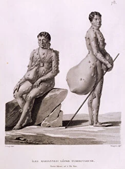
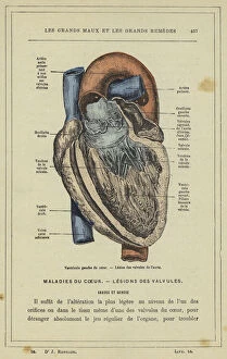
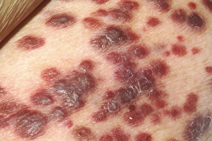
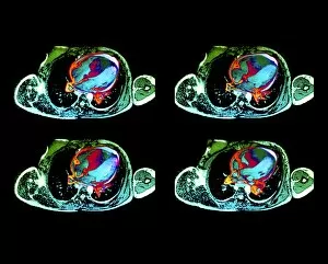
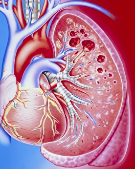
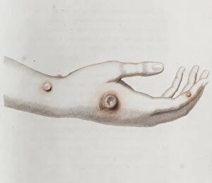
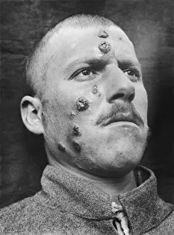
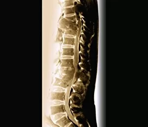
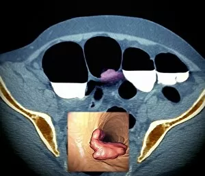
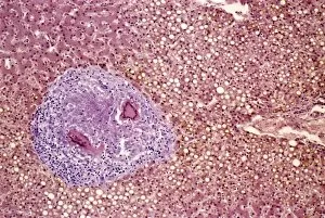
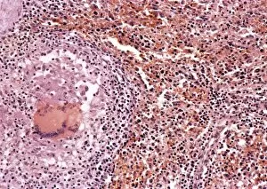
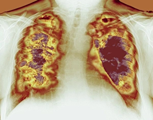
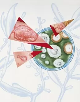
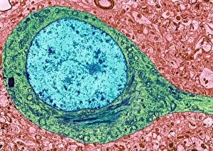
"Exploring the Varied Faces of Lesions: From Tuberculosis in Northern Mariana Islands to Fibroid Tumors in Female Uterus" In a remote corner of the Northern Mariana Islands, two men battle against tuberculosis, a disease that leaves its mark on their bodies through painful lesions. A striking colour lithograph reveals the intricate details of a lesion on the valvules de l'aorte, located within the left ventricle of the heart. On Guam, a man bravely faces elephantiasis, showcasing an extreme case captured during an expedition chronicled in Voyage Autour du Monde sur les Corvettes de. Coloured depictions showcase lesions both on arms and hands, shedding light on different manifestations and locations of these skin abnormalities. Delving into medical research, histopathology and pathophysiology studies unravel the complexities behind diabetic foot ulcers – debilitating lesions affecting individuals with diabetes. Psoriasis takes center stage as we explore this chronic autoimmune condition characterized by red patches covered with silvery scales – yet another form impacting millions worldwide. The devastating impact of AIDS is revealed through Kaposi's sarcoma lesions found on the skin of affected patients – highlighting how this disease affects not only internal organs but also external appearances. Cutting-edge MRI scans expose cardiac lymphoma lesions lurking within unsuspecting hearts – emphasizing early detection for improved treatment outcomes. Artistic representations bring pulmonary tuberculosis to life by illustrating lung cavities caused by this infectious disease that has plagued humanity throughout history. Peering into female anatomy uncovers fibroid tumors' presence within uteruses – underscoring how these benign growths can cause discomfort and complications for many women globally. A large eroded plaque adorned with areas of crust serves as a visual reminder that even seemingly innocuous skin conditions can evolve into significant lesions requiring medical attention.