
Light Sensitive Collection
"Capturing the Essence of Light: Exploring the Intricacies of Light-Sensitive Cells" In our quest to understand the wonders of vision


All Professionally Made to Order for Quick Shipping
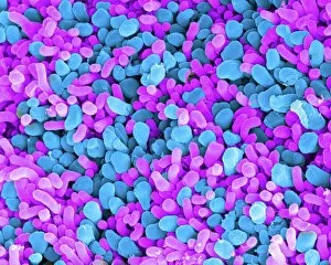
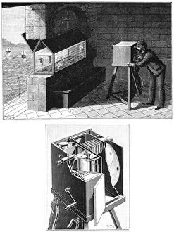
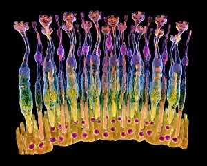
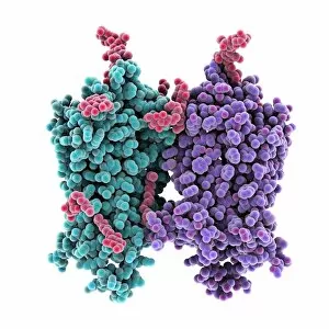
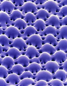
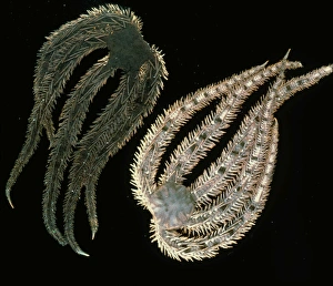
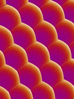
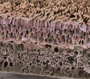
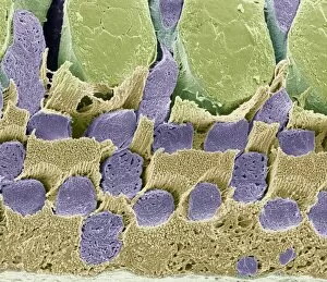
"Capturing the Essence of Light: Exploring the Intricacies of Light-Sensitive Cells" In our quest to understand the wonders of vision, we delve into the fascinating world of light-sensitive cells. Meet the rod and cone cells, guardians of our sight. SEM C014 / 4866 and SEM C014 / 4864 images reveal their intricate structures, showcasing their vital role in transmitting visual information to our brain. But humans aren't alone in possessing such remarkable abilities. Behold the mesmerizing Holothuria impatiens, a light-sensitive sea cucumber that astounds us with its defensive cuvierian respiratory tubules extruding when exposed to light. Nature's own defense mechanism at work. Taking a leap forward in time, we encounter Etienne Jules Marey's groundbreaking invention – the chambre chrono-photographique. This pioneering cine-camera paved the way for capturing motion pictures as we know them today. A testament to humanity's relentless pursuit of understanding and harnessing light. Zooming back into our eyes' inner workings, F008 / 0713 image reveals an eye retina brimming with life-sustaining photoreceptor cells. Rods and cones (F008 / 0719) work harmoniously within this delicate network to perceive colors, shapes, and depth perception. The eye retina never ceases to amaze us; F008 / 0714 showcases its intricate patterns while F008 / 0712 highlights its resilience against external factors. Every detail matters when it comes to preserving our precious gift of sight. As we explore further through F008/0715-F008/0718 images, we witness how these retinal layers adapt seamlessly between darkness and brightness – constantly adjusting sensitivity levels for optimal vision. In this captivating journey through light-sensitive wonders, let us appreciate both nature's ingenuity and human innovation as they converge upon one common goal: unraveling the mysteries hidden within the realm of light.