
Lung Collection
"Lungs: The Vital Organ for Breathing and Beyond" The lungs, a crucial component of the respiratory system, play a pivotal role in sustaining human life


All Professionally Made to Order for Quick Shipping
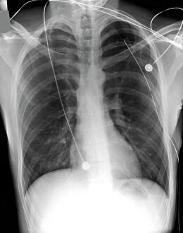
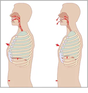
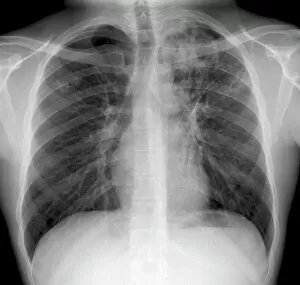
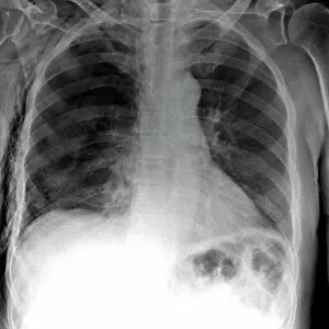
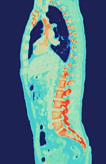
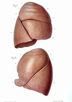
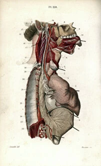
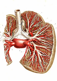
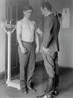
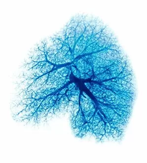
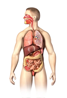
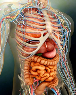
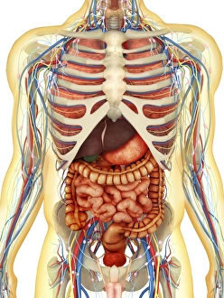
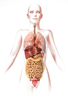
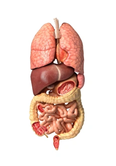
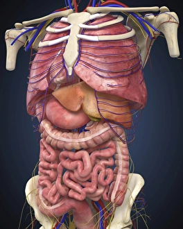
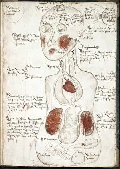
"Lungs: The Vital Organ for Breathing and Beyond" The lungs, a crucial component of the respiratory system, play a pivotal role in sustaining human life. From treating pneumothorax to understanding comparative embryology, these remarkable organs have captivated medical professionals throughout history. In 1932, the Department of Comparative and Human Anatomy at the American Museum of Natural History unveiled a groundbreaking chart showcasing comparative embryology from fish to man. This visual masterpiece shed light on the evolutionary journey that led to our complex lung structure. X-rays have been instrumental in diagnosing various lung conditions. They aid in identifying pneumothorax treatment options and detecting tuberculosis. These images provide valuable insights into the mechanics of respiration and help doctors understand how diseases affect this intricate organ system. Delving deeper into anatomy, bronchus and bronchial tubes are essential components within our lungs' framework. Understanding their structure enables healthcare professionals to diagnose conditions like tension pneumothorax accurately. Cystic fibrosis is another condition that affects lung health significantly. This genetic disorder leads to thick mucus accumulation within airways, causing breathing difficulties. Researchers tirelessly work towards finding effective treatments for this challenging disease. Konstantin Buteyko, a Soviet doctor renowned for his innovative breathing techniques, revolutionized respiratory therapy by emphasizing nasal breathing over mouth-breathing practices. His methods continue to inspire modern approaches in managing various lung disorders effectively. Modern imaging technologies such as MRI scans provide detailed visuals of normal torsos with healthy lungs or those affected by ailments requiring medical attention. These non-invasive procedures offer invaluable information for accurate diagnosis and treatment planning. Advertisements promoting cold mixtures often highlight their efficacy in relieving chest congestion caused by common colds or flu-like symptoms affecting our precious lungs temporarily. Human lungs themselves are marvels of engineering - delicate yet resilient enough to withstand constant exposure to environmental pollutants while facilitating efficient gas exchange necessary for survival. From historical breakthroughs to cutting-edge advancements, the study of lungs remains a fascinating field.