
Lymphocytes Collection
"Lymphocytes: The Mighty Warriors of our Immune System" Lymphocytes, the unsung heroes of our immune system


All Professionally Made to Order for Quick Shipping
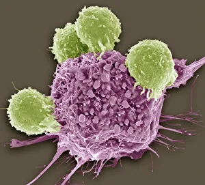
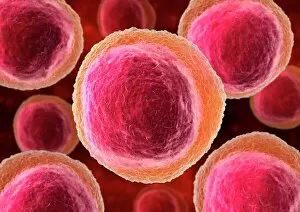
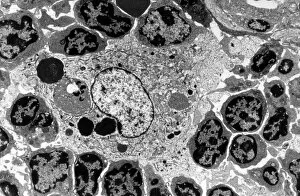
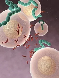
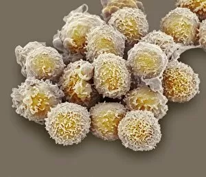
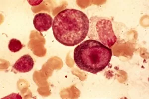
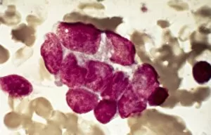
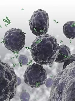
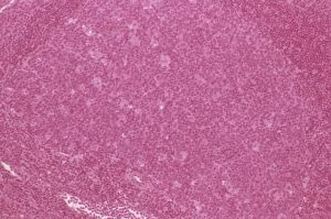
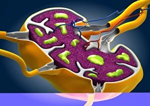
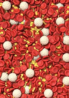
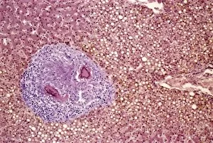
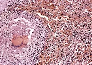
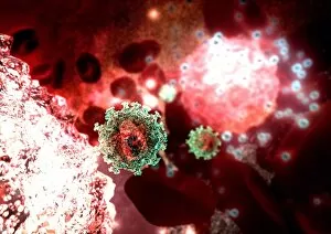
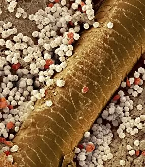
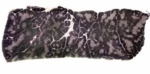
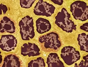
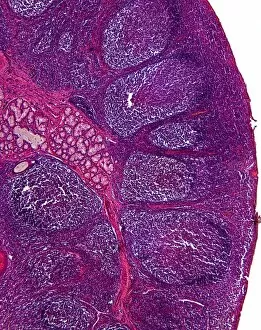
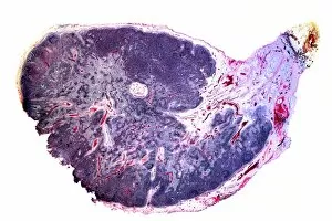
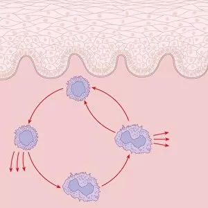
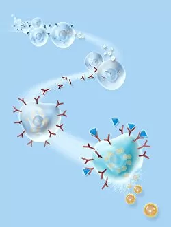
"Lymphocytes: The Mighty Warriors of our Immune System" Lymphocytes, the unsung heroes of our immune system, play a crucial role in defending our bodies against various diseases and infections. These remarkable white blood cells come in different forms, each with its own unique function. T lymphocytes (T cells) are specialized warriors that target cancer cells head-on. In SEM C001/1679, we witness their relentless pursuit to eliminate these malignant invaders. Their extraordinary ability to recognize abnormal cells makes them invaluable allies in the fight against cancer. In an artistic representation, lymphocyte white blood cells are showcased diligently patrolling through our bloodstream like vigilant guardians. They work hand-in-hand with macrophages as seen in TEM images, forming a formidable team that engulfs and destroys harmful pathogens. SEM images reveal another fascinating aspect - their presence within hair follicles. This surprising discovery suggests that they may have a vital role beyond just immunity. A cutaway view of a Lymph Node highlights the diverse tissue types where lymphocytes reside and interact with other immune components. This intricate network ensures efficient communication and coordination among various defense mechanisms. NK (Natural Killer) cells exhibit their exceptional abilities by attacking cancerous cells relentlessly as depicted in awe-inspiring artwork. Their precision strikes make them potent weapons against tumors. Antibodies join forces with bacteria-fighting lymphocytes in captivating artwork showcasing their symbiotic relationship. Together, they neutralize harmful microbes and safeguard our well-being. The Thymus gland takes center stage under light micrograph C015/4970 revealing its importance as the birthplace for T-cells maturation – shaping them into powerful defenders ready to combat threats effectively. SEM images C016/9140 & C016/9138 provide breathtaking close-ups of lymphocyte white blood cells displaying their intricate structures up-close; reminding us how nature's design is truly awe-inspiring at microscopic levels. However, not all lymphocyte stories have a happy ending.