
Lysis Collection
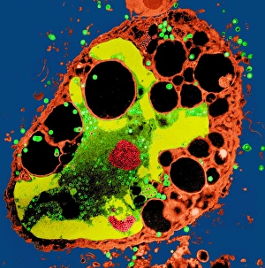
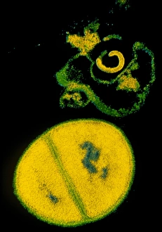
"Unveiling the Battle Within: Exploring Lysis in Viral Infections" Witness the microscopic battlefield as a cell infected with herpes virus succumbs to lysis


All Professionally Made to Order for Quick Shipping
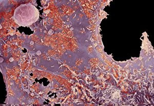
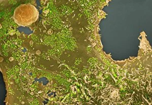
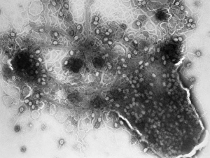
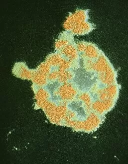
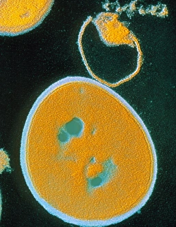
"Unveiling the Battle Within: Exploring Lysis in Viral Infections" Witness the microscopic battlefield as a cell infected with herpes virus succumbs to lysis, revealing the destructive power of this viral invader. (SEM C014 / 0604) Dive into the intricate world of cellular warfare as another herpes-infected cell undergoes lysis, highlighting the relentless nature of this viral infection. (SEM C014 / 0602) Explore the aftermath of a fierce battle between a herpes virus and its host cell, where lysis becomes an inevitable outcome, leaving behind only remnants of what was once a thriving organism. (SEM C014 / 0600) Behold the captivating imagery as yet another cell falls victim to herpes virus-induced lysis, showcasing both the beauty and devastation that occurs at such minuscule scales. (SEM C014 / 0603) Delve deeper into the realm of viral infections with an up-close look at a cell ravaged by herpes simplex virus under TEM imaging – witness firsthand how these tiny invaders dismantle their host from within. Uncover Cholera cytolysin's deadly secret as it unleashes its potent toxin upon unsuspecting cells – observe its devastating effects through SEM imaging for an eye-opening experience. (C015 / 6228) Step into a world where bacterial hosts fall prey to bacteriophage viruses - witness E. coli's struggle against T4 phages under TEM imaging, capturing moments before complete bacterial lysis occurs. Marvel at nature's microscopic assassins - bacteriophage viruses - as they relentlessly target and infect bacteria in their quest for survival and replication. Explore TEM imagery revealing bacterial cells succumbing to T4 phage infection-induced lysis; providing valuable insights into how these viruses exploit their hosts' resources for propagation.