Fraud Blocker Skip to main content

Macromolecule Collection (page 4)

Macromolecules, the building blocks of life, are at the forefront of scientific innovation

Background imageMacromolecule Collection: Chromosome segregation protein

Chromosome segregation protein, molecular model. This proteins function is to aid the process of chromosome segregation during cell division and replication

Background imageMacromolecule Collection: Nerve growth factor protein complex

Nerve growth factor protein complex, molecular model. This complex consists of nerve growth factor (NGF) in complex with four binding proteins

Background imageMacromolecule Collection: Thymidylic acid-ribonuclease A complex

Thymidylic acid-ribonuclease A complex. Molecular model of a thymidylic acid tetramer (blue) in complex with ribonuclease A (red)

Background imageMacromolecule Collection: DNA bundle on silicon nanopillars, SEM

DNA bundle on silicon nanopillars, SEM
DNA bundle on silicon nanopillars. Scanning electron micrograph (SEM) of a DNA (deoxyribonucleic acid) bundle and silicon nanopillars used to obtain the first high-contrast direct images of DNA

Background imageMacromolecule Collection: Exosome complex, molecular model

Exosome complex, molecular model. This multi-protein complex functions to break up strands of RNA (ribonucleic acid, pink) during biochemical processes

Background imageMacromolecule Collection: MyoD muscle protein-DNA complex

MyoD muscle protein-DNA complex. Molecular model of the MyoD muscle protein (green and blue) bound to a strand of DNA (deoxyribonucleic acid, orange and green)

Background imageMacromolecule Collection: Bacterial ribosome and protein synthesis

Bacterial ribosome and protein synthesis. Molecular model showing a bacterial ribosome reading an mRNA (messenger ribonucleic acid) strand (blue) and synthesising a protein

Background imageMacromolecule Collection: Canine parvovirus capsid C015 / 8460

Canine parvovirus capsid C015 / 8460
Canine parvovirus capsid, molecular model. Canine parvovirus type 2 (CPV2) was first recognized in 1978 and spread worldwide in less than two years

Background imageMacromolecule Collection: VDAC-1 ion channel protein C015 / 8250

VDAC-1 ion channel protein C015 / 8250
VDAC-1 ion channel protein, molecular model. This is the human voltage-dependent anion-selective channel protein 1 (VDAC-1)

Background imageMacromolecule Collection: Streptavidin bacterial protein C015 / 8458

Streptavidin bacterial protein C015 / 8458
Streptavidin bacterial protein, molecular model. Streptavidin is a protein obtained from the bacterium Streptomyces avidinii

Background imageMacromolecule Collection: Calcium ATPase ion pump C015 / 7143

Calcium ATPase ion pump C015 / 7143
Calcium ATPase ion pump, molecular model. This enzyme is found in muscle cell membranes, where it pumps calcium in and out of muscle cells and controls muscle contractions

Background imageMacromolecule Collection: DNA repair protein AlkB with DNA C016 / 0547

DNA repair protein AlkB with DNA C016 / 0547
DNA repair protein AlkB with DNA. Molecular model of the DNA (deoxyribonucleic acid) repair protein AlkB (blue) bound to a strand of double-stranded DNA (ds-DNA, pink and yellow)

Background imageMacromolecule Collection: Sodium-potassium ion pump proteins C015 / 9993

Sodium-potassium ion pump proteins C015 / 9993
Sodium-potassium ion pump proteins, molecular model. Sodium-potassium ATPase (adenosine triphosphatase) is an ATP-powered ion pump found in all animal cells

Background imageMacromolecule Collection: Thrombin complexed with fibrinogen C015 / 7148

Thrombin complexed with fibrinogen C015 / 7148
Thrombin complexed with fibrinogen, molecular model. The thrombin molecules (left and right, purple and green) are bound to the central part of the fibrinogen molecule (centre, multiple colours)

Background imageMacromolecule Collection: 3-hydroxyacyl-CoA dehydrogenase C015 / 9940

3-hydroxyacyl-CoA dehydrogenase C015 / 9940
3-hydroxyacyl-CoA dehydrogenase, molecular model. This enzyme is found in human heart tissue, and catalyzes a reaction that is part of the beta-oxidation pathway

Background imageMacromolecule Collection: Streptavidin bacterial protein C015 / 8457

Streptavidin bacterial protein C015 / 8457
Streptavidin bacterial protein, molecular model. Streptavidin is a protein obtained from the bacterium Streptomyces avidinii

Background imageMacromolecule Collection: Sodium-potassium ion pump proteins C015 / 9997

Sodium-potassium ion pump proteins C015 / 9997
Sodium-potassium ion pump proteins, molecular model. Sodium-potassium ATPase (adenosine triphosphatase) is an ATP-powered ion pump found in all animal cells

Background imageMacromolecule Collection: Retinal-producing oxygenase enzyme C015 / 7150

Retinal-producing oxygenase enzyme C015 / 7150
Retinal-producing oxygenase enzyme, molecular model. This enzyme, an oxygenase, plays a role in the production and metabolism of retinal and other apocarotenoids

Background imageMacromolecule Collection: Central glycolytic gene regulator protein C016 / 0549

Central glycolytic gene regulator protein C016 / 0549
Central glycolytic gene regulator (CGGR) protein, molecular model. This protein binds to DNA (deoxyribonucleic acid) in the absence of glucose, blocking the transcription of certain genes

Background imageMacromolecule Collection: Haemoglobin, molecular model C015 / 9391

Haemoglobin, molecular model C015 / 9391
Haemoglobin, molecular model. This is deoxyhaemoglobin, the molecule in its non-oxygen bound state. Haemoglobin transports oxygen around the body in red blood cells

Background imageMacromolecule Collection: Hemolysin-coregulated protein

Hemolysin-coregulated protein, molecular model. This protein is from the bacterium Pseudomonas aeruginosa. It is a hexameric ring structure, named hcp1

Background imageMacromolecule Collection: Canine parvovirus capsid C015 / 8459

Canine parvovirus capsid C015 / 8459
Canine parvovirus capsid, molecular model. Canine parvovirus type 2 (CPV2) was first recognized in 1978 and spread worldwide in less than two years

Background imageMacromolecule Collection: DNA repair protein AlkB with DNA C016 / 0546

DNA repair protein AlkB with DNA C016 / 0546
DNA repair protein AlkB with DNA. Molecular model of the DNA (deoxyribonucleic acid) repair protein AlkB (purple) bound to a strand of double-stranded DNA (ds-DNA, red and green)

Background imageMacromolecule Collection: Interferon-DNA transcription complex C015 / 8251

Interferon-DNA transcription complex C015 / 8251
Interferon-DNA transcription complex, molecular model. Bound to the DNA (deoxyribonucleic acid, green and yellow) is transcription factor p65, interferon regulatory factor 7

Background imageMacromolecule Collection: Interferon-DNA transcription complex C015 / 8252

Interferon-DNA transcription complex C015 / 8252
Interferon-DNA transcription complex, molecular model. Bound to the DNA (deoxyribonucleic acid, pink and white) is transcription factor p65, interferon regulatory factor 7, interferon fusion protein

Background imageMacromolecule Collection: Vitamin B12 import proteins C015 / 9942

Vitamin B12 import proteins C015 / 9942
Vitamin B12 import proteins, molecular model. This complex is the import proteins btuC, btuD, and btuF. The first two together form BtuCD

Background imageMacromolecule Collection: VDAC-1 ion channel protein C015 / 8249

VDAC-1 ion channel protein C015 / 8249
VDAC-1 ion channel protein, molecular model. This is the human voltage-dependent anion-selective channel protein 1 (VDAC-1)

Background imageMacromolecule Collection: Thrombin complexed with fibrinogen C015 / 7149

Thrombin complexed with fibrinogen C015 / 7149
Thrombin complexed with fibrinogen, molecular model. The thrombin molecules (left and right, brown and pink) are bound to the central part of the fibrinogen molecule (centre, multiple colours)

Background imageMacromolecule Collection: Simian virus (SV40) large T antigen C015 / 7069

Simian virus (SV40) large T antigen C015 / 7069
Simian virus (SV40) large T antigen, molecular model. This antigen is from the simian vacuolating virus 40 (SV40). Large T antigens play a role in regulating the viral life cycle of

Background imageMacromolecule Collection: Haemoglobin, molecular model C015 / 9392

Haemoglobin, molecular model C015 / 9392
Haemoglobin, molecular model. This is deoxyhaemoglobin, the molecule in its non-oxygen bound state. Haemoglobin transports oxygen around the body in red blood cells

Background imageMacromolecule Collection: 3-hydroxyacyl-CoA dehydrogenase C015 / 9941

3-hydroxyacyl-CoA dehydrogenase C015 / 9941
3-hydroxyacyl-CoA dehydrogenase, molecular model. This enzyme is found in human heart tissue, and catalyzes a reaction that is part of the beta-oxidation pathway

Background imageMacromolecule Collection: Vitamin B12 import proteins C015 / 9943

Vitamin B12 import proteins C015 / 9943
Vitamin B12 import proteins, molecular model. This complex is the import proteins btuC, btuD, and btuF. The first two together form BtuCD

Background imageMacromolecule Collection: Calcium ATPase ion pump C015 / 7142

Calcium ATPase ion pump C015 / 7142
Calcium ATPase ion pump, molecular model. This enzyme is found in muscle cell membranes, where it pumps calcium in and out of muscle cells and controls muscle contractions

Background imageMacromolecule Collection: Retinal-producing oxygenase enzyme C015 / 7151

Retinal-producing oxygenase enzyme C015 / 7151
Retinal-producing oxygenase enzyme, molecular model. This enzyme, an oxygenase, plays a role in the production and metabolism of retinal and other apocarotenoids

Background imageMacromolecule Collection: Adeno-associated virus, molecular model C018 / 0449

Adeno-associated virus, molecular model C018 / 0449
Adeno-associated virus (aV), molecular model. This image shows the core protein of the non-enveloped virus that surrounds the genetic material

Background imageMacromolecule Collection: Hepatitis B virus, molecular model C018 / 0455

Hepatitis B virus, molecular model C018 / 0455
Hepatitis B virus core protein, molecular model. The virus causes hepatitis B, an inflammatory liver disease. The core proteins enclose the virus DNA and are in turn surrounded by a lipid envelope

Background imageMacromolecule Collection: Norovirus capsid, molecular model C018 / 0457

Norovirus capsid, molecular model C018 / 0457
Norovirus capsid, molecular model. Also known as the winter vomiting bug, Noroviruses cause gastroenteritis and are highly contagious, infecting approximately 267 million people a year

Background imageMacromolecule Collection: Astrovirus capsid, molecular model C018 / 0450

Astrovirus capsid, molecular model C018 / 0450
Astrovirus capsid, molecular model. This icosahedral virus was identified in 1975 using electron microscopy. It has a characteristic five-pointed symmetry to its surface, as seen here

Background imageMacromolecule Collection: Hepatitis E virus, molecular model C018 / 0445

Hepatitis E virus, molecular model C018 / 0445
Hepatitis E virus core protein, molecular model. The virus causes hepatitis E, an inflammatory liver disease that usually only lasts a few weeks

Background imageMacromolecule Collection: Human polio virus, molecular model

Human polio virus, molecular model
Human polio virus capsid, molecular model. Poliovirus causes poliomyelitis, a disease that can cause paralysis in up to 2 percent of patients, and in some cases death

Background imageMacromolecule Collection: Clathrin lattice, molecular model C018 / 0453

Clathrin lattice, molecular model C018 / 0453
Clathrin lattice, molecular model. This polyhedral protein lattice coats eukaryotic cell membranes (vesicles) and is involved in protein secretion and membrane trafficking

Background imageMacromolecule Collection: Clathrin lattice, molecular model C018 / 0452

Clathrin lattice, molecular model C018 / 0452
Clathrin lattice, molecular model. This polyhedral protein lattice coats eukaryotic cell membranes (vesicles) and is involved in protein secretion and membrane trafficking

Background imageMacromolecule Collection: Clathrin lattice, molecular model C018 / 0454

Clathrin lattice, molecular model C018 / 0454
Clathrin lattice, molecular model. This polyhedral protein lattice coats eukaryotic cell membranes (vesicles) and is involved in protein secretion and membrane trafficking

Background imageMacromolecule Collection: KSHV virus capsid, molecular model C018 / 0456

KSHV virus capsid, molecular model C018 / 0456
KSHV virus capsid, molecular model. KSHV is Kaposis sarcoma-associated herpesvirus. The virus is an oncovirus, which is a virus that can cause cancer

Background imageMacromolecule Collection: Ricin molecule, artwork C017 / 3656

Ricin molecule, artwork C017 / 3656
Ricin molecule. Computer artwork showing the structure of a molecule of the toxic protein ricin. Ricin comprises two entwined amino acid chains; A (yellow) and B (blue)

Background imageMacromolecule Collection: Ricin molecule, artwork C017 / 3655

Ricin molecule, artwork C017 / 3655
Ricin molecule. Computer artwork showing the structure of a molecule of the toxic protein ricin. Ricin comprises two entwined amino acid chains; A (yellow) and B (blue)

Background imageMacromolecule Collection: SV40 virus capsid, molecular model

SV40 virus capsid, molecular model. Simian virus 40 (SV40) is found in monkeys such as Rhesus monkeys and macaques. Potentially tumour-causing, it is used in laboratory research and in vaccines

Background imageMacromolecule Collection: Dengue virus capsid, molecular model

Dengue virus capsid, molecular model. This virus, transmitted by mosquito bites, causes the tropical disease dengue fever in humans


All Professionally Made to Order for Quick Shipping

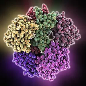
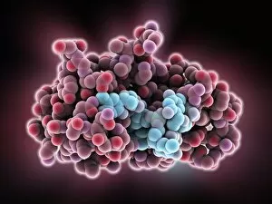
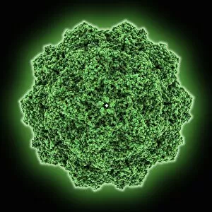
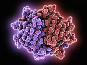
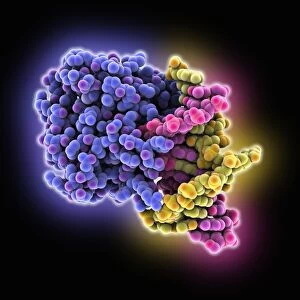
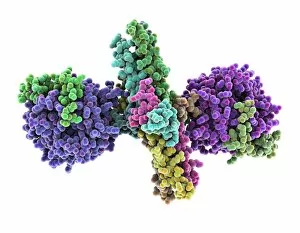
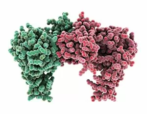
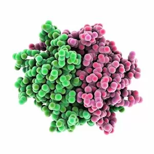
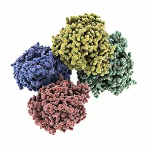
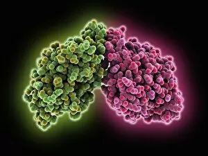
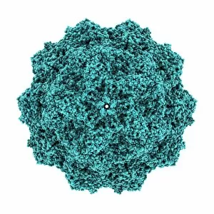
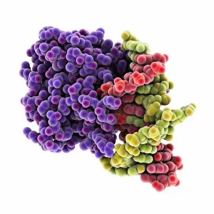
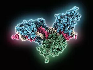
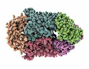
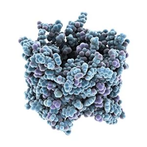
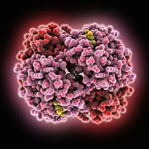
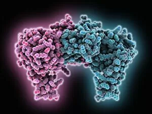
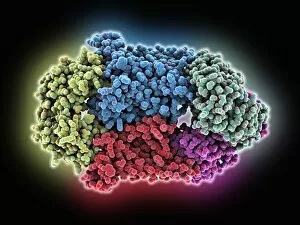
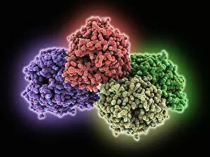
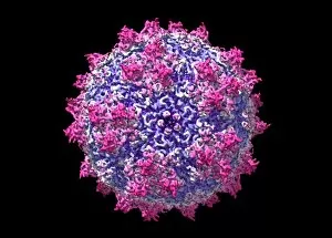
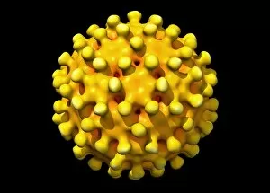
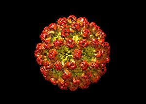
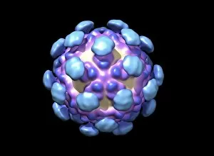
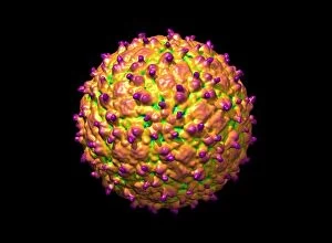
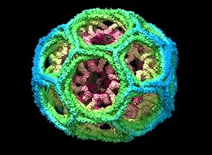
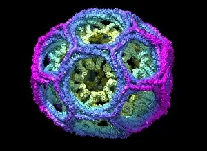
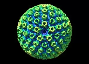
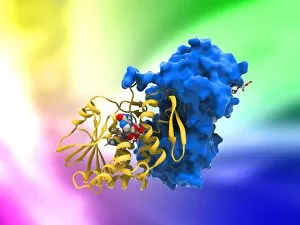
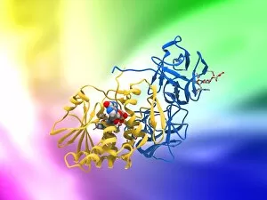
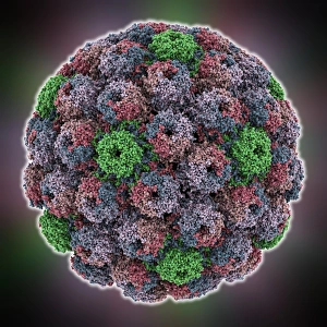
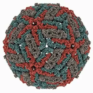
Macromolecules, the building blocks of life, are at the forefront of scientific innovation. Nanotube technology has revolutionized various fields, enabling advancements in medicine and electronics. In this captivating computer artwork, we witness the intricate Zinc fingers binding to a DNA strand, showcasing their crucial role in gene regulation. Carbon nanotubes have also emerged as remarkable materials with immense potential. Their unique structure and properties make them ideal for applications ranging from energy storage to drug delivery systems. Computer-generated images depict these carbon nanotubes in all their glory. The SARS coronavirus protein is another macromolecule that has garnered significant attention due to its role in viral infection. Scientists tirelessly study it to develop effective treatments against deadly outbreaks. Computer models allow us to explore complex structures like Bacteriophage phi29—a virus that infects bacteria—providing insights into its mechanisms and aiding in the development of targeted therapies. Simian immunodeficiency virus (SIV), closely related to HIV, poses a global health challenge. Understanding its macromolecular components helps researchers devise strategies for prevention and treatment. Rhodopsin protein molecule captures our imagination with its vital function in vision. Its elegant structure enables light detection and initiates visual signals within our eyes. TFAM transcription factor bound to DNA C015/7059 showcases how macromolecules regulate gene expression by interacting with specific regions on DNA strands—an essential process for cell functioning and development. These glimpses into the world of macromolecules highlight their significance across diverse disciplines—from cutting-edge technologies like nanotube engineering to unraveling infectious diseases or understanding fundamental biological processes. As scientists continue exploring these fascinating molecules, they pave the way for groundbreaking discoveries that shape our future.