
Malignancy Collection
"Malignancy: Unveiling the Battle Within" In this captivating journey through the realm of malignancy


All Professionally Made to Order for Quick Shipping
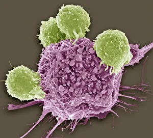
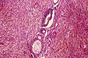
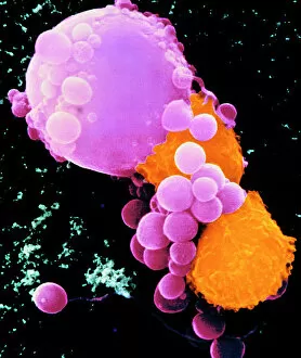
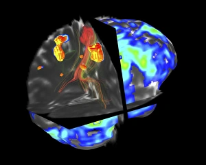
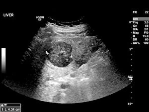
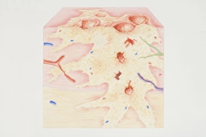
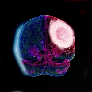
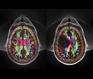
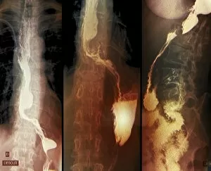
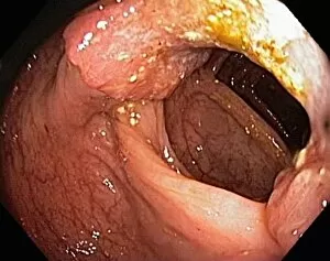
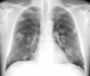
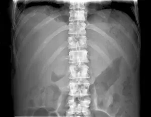
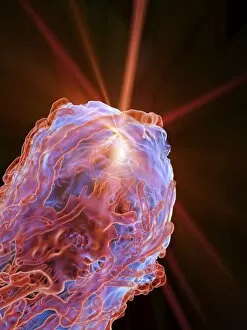
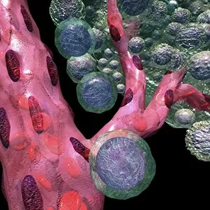
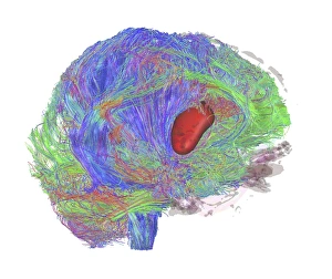
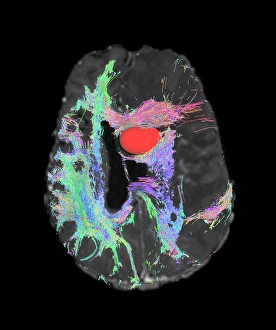
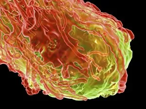
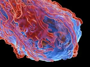
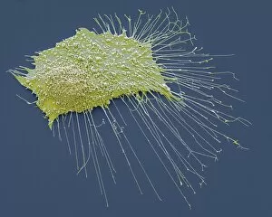
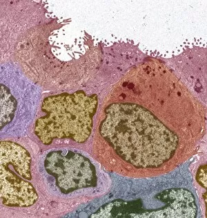
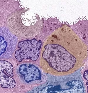
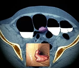
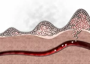
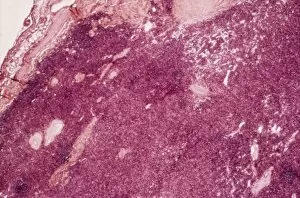
"Malignancy: Unveiling the Battle Within" In this captivating journey through the realm of malignancy, we witness the relentless struggle between T lymphocytes and cancer cells. A mesmerizing SEM image (C001 / 1679) reveals a colorful spectacle as lymphocytes valiantly attack a cancer cell, embodying hope in the face of adversity. Moving deeper into this intricate world, an ovarian cancer light micrograph (C015 / 7103) unveils the hidden intricacies of this disease. The delicate brushstrokes of an oil painting depicting breast cancer (C017 / 1273) evoke both beauty and pain, reminding us that art can capture even the most complex emotions. Venturing into the depths of our minds, brain tumors come to life through fMRI and tractography scans (C017 / 7102). These images showcase not only their physical presence but also their impact on neural pathways. DTI modeling (C017 / 7060) further unravels these enigmatic growths while three-dimensional CT scans (C016 / 6414) provide a comprehensive view from all angles. The battle against malignancy extends beyond just one organ or system. Throat cancer X-rays and secondary lung cancers X-ray serve as stark reminders that no part is immune to its reach. An ultrasound scan exposes secondary liver cancer's insidious invasion (C017/7764), urging us to remain vigilant in our fight against this formidable foe. A cross-section diagram portrays a vivid snapshot of a malignant tumor's complexity - calcium deposits, blood vessels, tumorous outgrowths - all entwined with nerve fibers amidst ulcerated areas and bleeding wounds. This visual representation serves as a reminder that behind every diagnosis lies an intricate web affecting multiple facets of our being. As science progresses hand-in-hand with compassion, we delve deeper into understanding malignancies' secrets.