Fraud Blocker Skip to main content

Mandible Collection

The mandible, also known as the jawbone, is a fascinating and crucial component of our skull anatomy

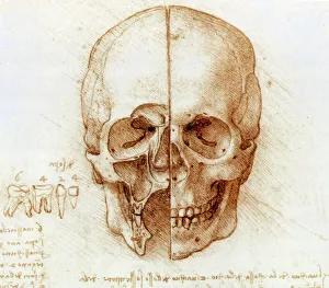
Background imageMandible Collection: Skull anatomy by Leonardo da Vinci

Skull anatomy by Leonardo da Vinci. Historical artwork and notes on the anatomy of the human skull and teeth, by the Italian artist and scientist Leonardo da Vinci (1452-1519)

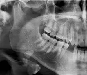
Background imageMandible Collection: Panoramic dental X-ray

Panoramic dental X-ray showing all the teeth in the upper and lower jaws

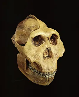
Background imageMandible Collection: Paranthropus boisei (Zinjanthropus) cranium (OH5)

Paranthropus boisei (Zinjanthropus) cranium (OH5)
Cast of the cranium of a young male of Paranthropus boisei discovered by Mary Leakey in 1959 at Olduvai Gorge. The specimen which is the Holotype of Zinjanthropus boisei (Leakey 1959)

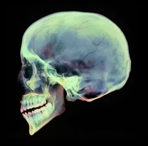
Background imageMandible Collection: Human skull, X-ray

Human skull, X-ray
Human skull. Coloured X-ray of a sagittal section through a human skull. The skull has been sliced in half down the centre, revealing details of the internal structures

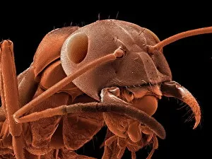
Background imageMandible Collection: Red-barbed ant, SEM

Red-barbed ant, SEM
Red-barbed ant (Formica rufibarbis), coloured scanning electron micrograph (SEM). This ant is common in mainland Europe, but rare in the UK

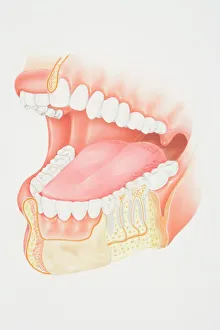
Background imageMandible Collection: Cross-section diagram of human mouth and jaw

Cross-section diagram of human mouth and jaw

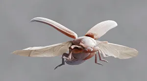
Background imageMandible Collection: Red flour beetle in flight

Red flour beetle in flight
Red flour beetle. Coloured scanning electron micrograph of a red flour beetle (Tribolium castaneum) in flight. One of the most significant pests of dried foods and the milling industry

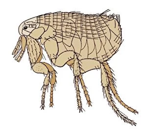
Background imageMandible Collection: Black Death rat flea, artwork

Black Death rat flea, artwork
Oriental rat flea (Xenopsylla cheopis), artwork. This flea was the primary vector of the bacterium Yersinia pestis, which caused the Black Death

Background imageMandible Collection: Fractured jawbone, X-ray

Fractured jawbone, X-ray
Fractured jawbone. Coloured X-ray of a human jaw showing a fractured lower jawbone (mandible). The fracture has occurred to the left side of the lower jawbone (bottom right, orange)

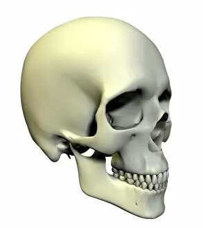
Background imageMandible Collection: Skull

Skull. Computer artwork of an oblique view of a human skull. The bones of the cranium are fused together to form a solid structure that encloses and protects the brain

Background imageMandible Collection: Spiny spider, SEM

Spiny spider, SEM
Spiny spider. Coloured scanning electron micrograph (SEM) of a spiny spider (Gasteracantha sp.), showing its characteristic spines (red) and large mouthparts (grey, centre left)
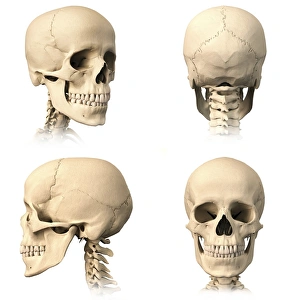
Background imageMandible Collection: Anatomy of human skull from different angles

Anatomy of human skull from different angles

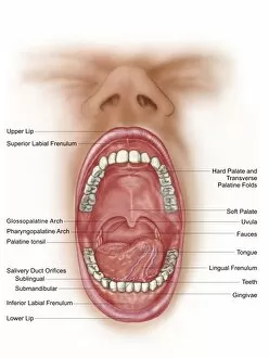
Background imageMandible Collection: Anatomy of human mouth cavity

Anatomy of human mouth cavity

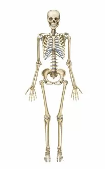
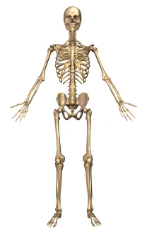
Background imageMandible Collection: Human skeletal system, front view

Human skeletal system, front view

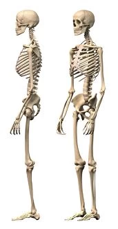
Background imageMandible Collection: Anatomy of male human skeleton, side view and perspective view

Anatomy of male human skeleton, side view and perspective view

Background imageMandible Collection: Egyptian skull

Egyptian skull. British Museum. London. United Kingdom
Background imageMandible Collection: DDE-90039441

DDE-90039441
Northern Cardinal (Cardinalis cardinalis) male with deformed beak. Date: 31/01/2013

Background imageMandible Collection: Lateral Diagram of the Bones of the Human Skull, 1906 (engraving)

Lateral Diagram of the Bones of the Human Skull, 1906 (engraving)
2622231 Lateral Diagram of the Bones of the Human Skull, 1906 (engraving) by German School, (20th century); Private Collection; (add.info)

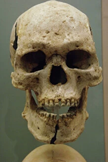
Background imageMandible Collection: Skull of a young girl. 18-20 years old. 3500-3400 BC

Skull of a young girl. 18-20 years old. 3500-3400 BC. Sigersdal Mose, Northern Zealand. National Museum of Denmark. Copenhagen. Denmark

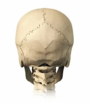
Background imageMandible Collection: Anatomy of human skull, rear view

Anatomy of human skull, rear view

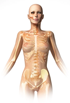
Background imageMandible Collection: Female body with bone skeleton and internal organs superimposed

Female body with bone skeleton and internal organs superimposed
Anatomy of female body with bone skeleton and all internal organs superimposed

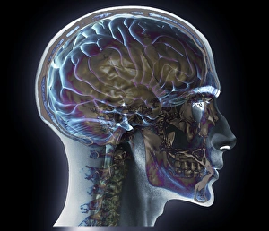
Background imageMandible Collection: Male human head with skull in ghost effect, side view

Male human head with skull in ghost effect, side view. on white background

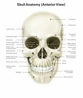
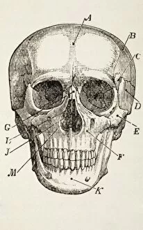
Background imageMandible Collection: Anterior view of human skull, with labels

Anterior view of human skull, with labels

Background imageMandible Collection: Base of human skull, inferior view, with labels

Base of human skull, inferior view, with labels

Background imageMandible Collection: Anatomy of human skull, cutaway view with half brain showing

Anatomy of human skull, cutaway view with half brain showing, front view

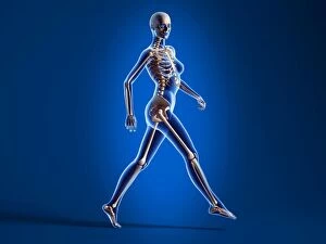
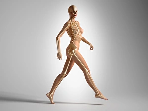
Background imageMandible Collection: X-ray view of a naked woman walking, with skeletal bones superimposed

X-ray view of a naked woman walking, with skeletal bones superimposed

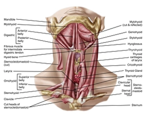
Background imageMandible Collection: Anatomy of human hyoid bone and muscles, anterior view

Anatomy of human hyoid bone and muscles, anterior view

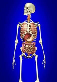
Background imageMandible Collection: Male skeleton with internal organs on blue background

Male skeleton with internal organs on blue background

Background imageMandible Collection: Male human skeleton in dynamic posture, front view

Male human skeleton in dynamic posture, front view

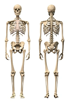
Background imageMandible Collection: Anatomy of male human skeleton, front view and back view

Anatomy of male human skeleton, front view and back view

Background imageMandible Collection: 3D rendering of a naked woman walking, with skeletal bones superimposed

3D rendering of a naked woman walking, with skeletal bones superimposed

Background imageMandible Collection: Anatomy of a male human head, with half muscles and half skull

Anatomy of a male human head, with half muscles and half skull, front view

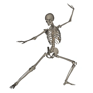
Background imageMandible Collection: Front view of human skeleton in fighting stance

Front view of human skeleton in fighting stance, isolated on white background

Background imageMandible Collection: Front view of a human skeleton posing

Front view of a human skeleton posing, isolated on white background

Background imageMandible Collection: Colored base of human skull, inferior view, with labels

Colored base of human skull, inferior view, with labels

Background imageMandible Collection: Salivary gland stone, X-ray

Salivary gland stone, X-ray
Salivary gland stone. X-ray of the jaw of a 36-year-old patient with a stone (white, lower left) in the right submaxillary gland

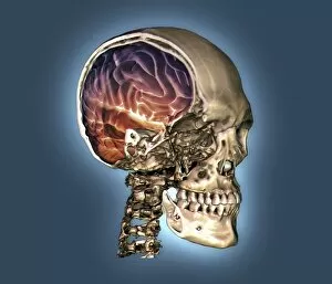
Background imageMandible Collection: Normal skull and brain, 3D CT scan C016 / 6333

Normal skull and brain, 3D CT scan C016 / 6333
Normal skull and brain. Coloured 3D computed tomography (CT) scan of the head of a 35 year old patient. Part of the skull has been cut-away to show the brain (purple and red)

Background imageMandible Collection: Normal head and neck, MRI and 3D CT scans C016 / 6337

Normal head and neck, MRI and 3D CT scans C016 / 6337
Normal head. Coloured composite image of a magnetic resonance imaging (MRI) scan of the brain and a 3D computed tomography (CT) scan of the head and neck of a 35 year old patient

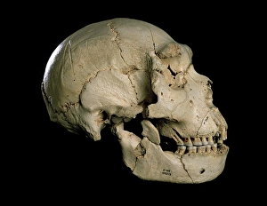
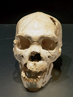
Background imageMandible Collection: Homo heidelbergensis skull (Cranium 5) C015 / 6921

Homo heidelbergensis skull (Cranium 5) C015 / 6921
Homo heidelbergensis skull (Cranium 5). Excavated in 1992 from the Sima de los Huesos pit in the Atapuerca foothills in Spain, this fossil skull dates from around 400, 000 years ago

Background imageMandible Collection: Rhyniognatha hirsti

Rhyniognatha hirsti
A pair of jaws (mandibles) preserved in a fragment of Rhynie Chert and mounted on a glass microscope slide. This is the worlds oldest fossil insect part

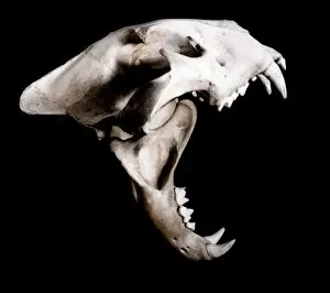
Background imageMandible Collection: Big cat skull

Big cat skull. Side (lateral) view of the skull of a big cat with its jaws wide open. The big cats, which include tigers and lions

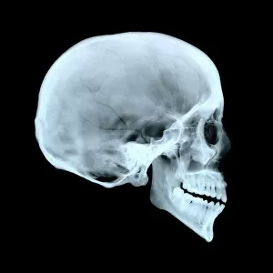
Background imageMandible Collection: Adult human skull

Adult human skull. Side view X-ray showing the cranium, eye socket, nasal area and teeth. For an X-ray of a babys skull see image P120/223 and for an X-ray of a childs skull see image number P120/224

Background imageMandible Collection: The Human Skull. From The Household Physician, Published Circa 1890

The Human Skull. From The Household Physician, Published Circa 1890

Background imageMandible Collection: Homo heidelbergensis. Skull number 5. Atapuerca, Spain

Homo heidelbergensis. Skull number 5. Atapuerca, Spain
Homo heidelbergensis. Skull number 5. Discovered in the Pit of Bones Site of Atapuerca (Spain). European Middle Pleistocene. Atapuerca. Spain

Background imageMandible Collection: Side view of human skull with parts exploded

Side view of human skull with parts exploded

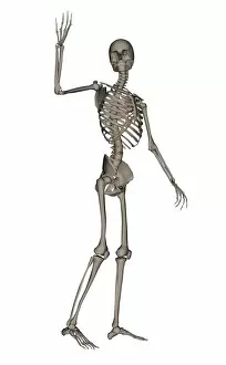
Background imageMandible Collection: Front view of human skeleton waving goodbye

Front view of human skeleton waving goodbye, isolated on white background

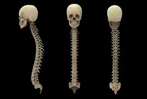
Background imageMandible Collection: 3D rendering of human vertebral column with skull

3D rendering of human vertebral column with skull

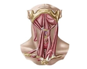
Background imageMandible Collection: Anatomy of human hyoid bone and neck muscles, anterior view

Anatomy of human hyoid bone and neck muscles, anterior view


All Professionally Made to Order for Quick Shipping

The mandible, also known as the jawbone, is a fascinating and crucial component of our skull anatomy. Leonardo da Vinci's detailed sketches in his "Skull Anatomy" showcase the intricacies of this bone. A panoramic dental X-ray allows us to examine the mandible's structure and its relationship with teeth. Looking back in time, the Paranthropus boisei (Zinjanthropus) cranium (OH5) provides valuable insights into our evolutionary history. The study of human skulls through X-rays reveals not only the mandible but also other vital structures within our head. Nature never fails to amaze us; even tiny creatures like the red-barbed ant can be observed under a scanning electron microscope (SEM), revealing their unique mandibles adapted for various tasks. Meanwhile, a cross-section diagram illustrates how our mouth and jaw work together harmoniously. Insects are no exception when it comes to showcasing intriguing mandibular adaptations. Witnessing a red flour beetle in flight or examining an artwork depicting a Black Death rat flea reminds us of nature's diversity and complexity. However, sometimes unfortunate incidents occur that affect this essential bone. Fractured jawbones captured by X-rays remind us of both medical challenges and advancements in treating such injuries. Zooming further into nature's wonders, we encounter spiny spiders under SEMs - their menacing-looking yet functional jaws ready for capturing prey. Ultimately, whether studying ancient fossils or observing modern-day organisms, understanding the mandible plays an integral role in comprehending head and neck anatomy as depicted beautifully through artworks dedicated to this subject matter.