
Maxillary Collection
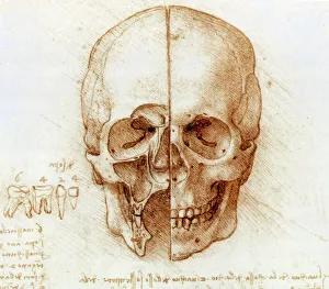
"Exploring the Intricacies of Maxillary: Unveiling the Secrets of Skull Anatomy by Leonardo da Vinci" Step into a world where art and science converge


All Professionally Made to Order for Quick Shipping
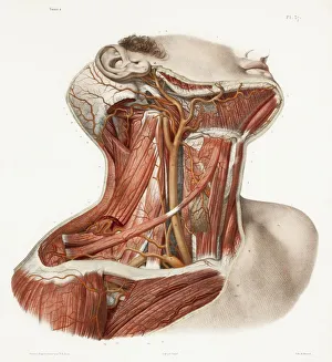
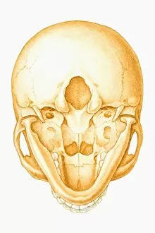
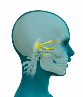
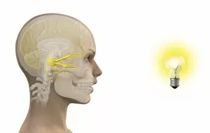
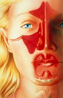
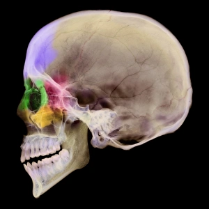
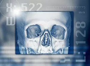
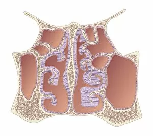
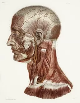
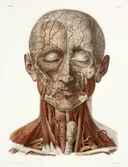
"Exploring the Intricacies of Maxillary: Unveiling the Secrets of Skull Anatomy by Leonardo da Vinci" Step into a world where art and science converge, as we delve into the fascinating realm of maxillary. In this captivating journey, we uncover hidden treasures within historical artwork and unravel the mysteries surrounding this vital part of our anatomy. Leonardo da Vinci, renowned for his artistic brilliance, also delved into skull anatomy. His meticulous illustrations provide us with a glimpse into the intricate structure of the maxillary region. Through his work, we gain a deeper understanding of its role in shaping our facial features. But it doesn't stop there; let's venture further into the depths. Exploring neck vascular anatomy reveals how blood vessels intertwine with maxillary nerves to ensure proper functioning. The olfactory and maxillary nerves play an essential role in transmitting sensory information from our nose to our brain – a testament to their significance. The fifth cranial nerve takes center stage as we examine its branches – specifically, the opthalmic and maxillary nerves. These intricate pathways are responsible for relaying sensations from various parts of our face, ensuring that every touch is felt and every pain is registered. An illustration showcasing a human skull seen from below offers a unique perspective on dental maxillary nerve regions. This visual representation allows us to comprehend their distribution throughout this complex area. Diving deeper still, we encounter different techniques employed in medical procedures such as infraorbital nerve blocks or anterior superior alveolar nerve blocks. These interventions target specific areas within the maxilla to alleviate pain or facilitate dental treatments - showcasing how knowledge about these nerves can be practically applied for patient care. As if that weren't intriguing enough, we stumble upon an unexpected phenomenon known as photic sneeze reflex – where exposure to bright light triggers uncontrollable sneezing episodes in some individuals due to cross-wiring between optic and trigeminal nerves.