
Micro Organism Collection
Microorganisms are the tiny, often unseen creatures that inhabit our world and play a crucial role in various ecosystems


All Professionally Made to Order for Quick Shipping
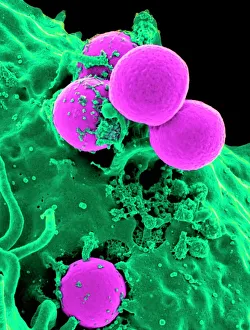
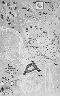
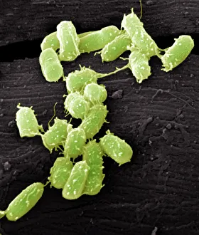
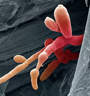
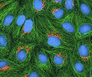
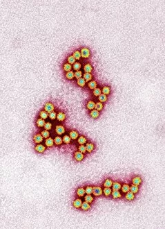
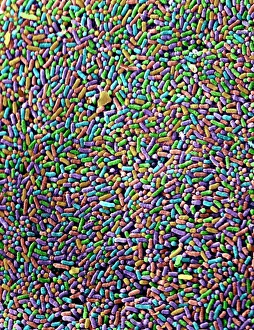
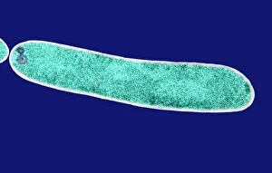
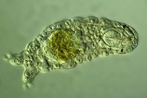
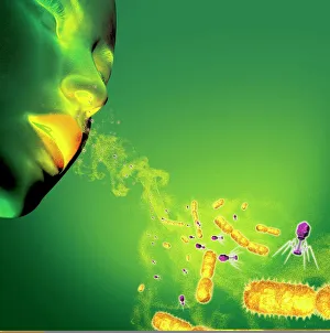
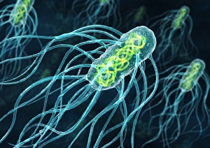
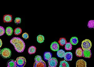
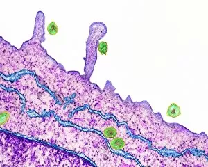
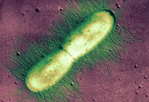
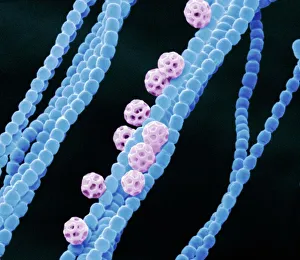
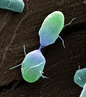
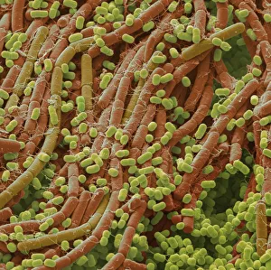
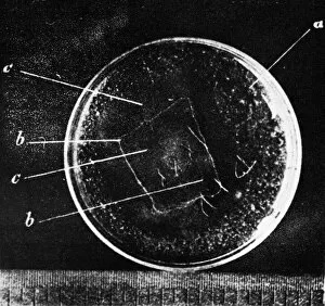
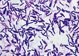
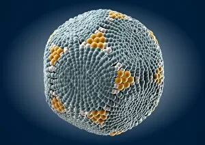
Microorganisms are the tiny, often unseen creatures that inhabit our world and play a crucial role in various ecosystems. From bacteria to fungi, they come in all shapes and sizes, as revealed by scientific imaging techniques. HeLa cells, immortalized human cells named after Henrietta Lacks, have been instrumental in medical research for decades. In a light micrograph labeled C017/8299, these remarkable cells can be seen dividing and multiplying under the lens of a microscope. Neutrophils are our body's first line of defense against bacterial infections. In an SEM image labeled C018/8596, we witness the incredible sight of a neutrophil engulfing MRSA (methicillin-resistant Staphylococcus aureus), showcasing the power of our immune system. E. Coli is a well-known bacterium that has both beneficial and harmful strains. In an SEM image, this bacterium appears like long rods with flagella propelling them forward. Candida fungus is another microscopic organism that can cause infections in humans. Under SEM magnification, it reveals its intricate branching structures resembling delicate webs or trees. Returning to HeLa cells but with a different perspective captured in light micrograph C017/8298; we observe their unique characteristics once again – their ability to grow rapidly and adapt to laboratory conditions while retaining essential cellular functions. Anthrax cultures depicted on historical diagrams remind us of the devastating impact certain microorganisms can have on society if weaponized or accidentally released into populations. Norovirus particles viewed through TEM show spherical viral bodies responsible for causing gastroenteritis outbreaks worldwide – highlighting how something so small can wreak havoc on our health. Salmonella bacteria appear rod-shaped under SEM magnification; these notorious culprits are known for causing food poisoning when ingested through contaminated food or water sources. Another view of E. coli at higher resolution using TEM reveals its intricate internal structure - providing insights into its mechanisms and potential targets for antibiotics.