Fraud Blocker Skip to main content

Microscope Collection

"Unveiling the Unseen: Exploring the Microscopic World" Step into a realm of hidden wonders

Background imageMicroscope Collection: Rosalind Franklin

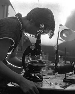
Rosalind Franklin
ROSALIND FRANKLIN Pioneer Molecular Biologist she made important contributions to understanding the structure of DNA

Background imageMicroscope Collection: False-coloured SEM (Scanning Electron Micrograph) of a Tardigrade (Tardigrada) amongst moss

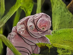
False-coloured SEM (Scanning Electron Micrograph) of a Tardigrade (Tardigrada) amongst moss, from Peak District National Park, Derbyshire, UK. This tardigrade is approximately 0.1 millimetres long

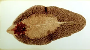
Background imageMicroscope Collection: Liver Fluke, microscope slide preparation of the whole animal

Liver Fluke, microscope slide preparation of the whole animal
PM-10632 Liver Fluke, microscope slide preparation of the whole animal

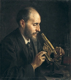
Background imageMicroscope Collection: RAMON Y CAJAL, Santiago (1852-1934). Spanish

RAMON Y CAJAL, Santiago (1852-1934). Spanish

Background imageMicroscope Collection: RAMON Y CAJAL, Santiago (1852-1934). Spanish

RAMON Y CAJAL, Santiago (1852-1934). Spanish doctor and histologist, Nobel Prize in 1906. Portrait in his laboratory

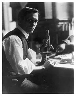
Background imageMicroscope Collection: Fleming / Photo / Microscope

Fleming / Photo / Microscope
SIR ALEXANDER FLEMING - Scottish bacteriologist at his desk with his microscope

Background imageMicroscope Collection: Fat cells, TEM

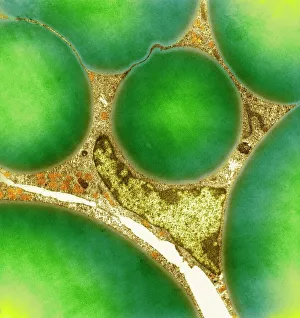
Fat cells, TEM
Fat cells. Coloured transmission electron micrograph (TEM) of a section through fat (adipose) tissue, consisting of fat-storing cells (adipocytes, green) supported by connective tissue

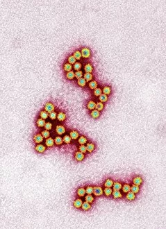
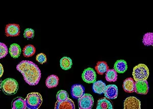
Background imageMicroscope Collection: Norovirus particles, TEM

Norovirus particles, TEM
Norovirus particles. Coloured transmission electron micrograph (TEM) of norovirus particles

Background imageMicroscope Collection: E. coli bacterium, TEM

E. coli bacterium, TEM
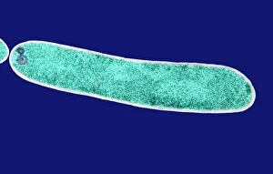
E. coli bacterium, coloured transmission electron micrograph (TEM). E. coli are Gram-negative rod-shaped bacteria that are part of the normal flora of the human gut

Background imageMicroscope Collection: Under the Microscope / C18

Under the Microscope / C18
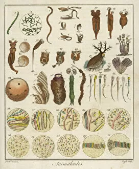
Animacules : microscopic creatures as seen under a microscope; the last two rows are human sperm

Background imageMicroscope Collection: Snail teeth

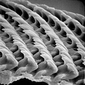
Snail teeth

Background imageMicroscope Collection: Lubbock / John / Graphic 84

Lubbock / John / Graphic 84
SIR JOHN LUBBOCK 1st BARON AVEBURY Liberal MP and writer of popular science books

Background imageMicroscope Collection: Plasmodium sp. malarial parasite

Plasmodium sp. malarial parasite
Scanning electron microscope image of a malarial protozoal parasite. The parasite requires the anopheles mosquito to complete its life cycle

Background imageMicroscope Collection: Plasma cells, TEM

Plasma cells, TEM
Plasma cells. Coloured transmission electron micrograph (TEM) of plasma cells. Plasma cells are mature B lymphocytes (white blood cells) that produce and secrete antibodies during an immune response

Background imageMicroscope Collection: Picture No. 10876997

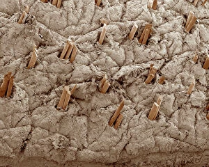
Picture No. 10876997
Scanning Electron Micrograph (SEM): Human Skin with Hair Follicles Date:

Background imageMicroscope Collection: Illustration of eye seeing through microscope

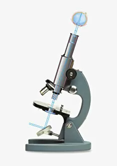
Illustration of eye seeing through microscope

Background imageMicroscope Collection: Picture No. 11014612

Picture No. 11014612
Quinidine Rippled Quinidine is a chemical precurser to the antimalarial drug quinine - The image was produced using a polarising microscope and post processed in Adobe Photoshop Date:

Background imageMicroscope Collection: Picture No. 10873704

Picture No. 10873704
Sleeping Sickness Parasite in red blood cells (Trypanosoma sp. ) Date:

Background imageMicroscope Collection: Nils and Aage Bohr in laboratory C016 / 8375

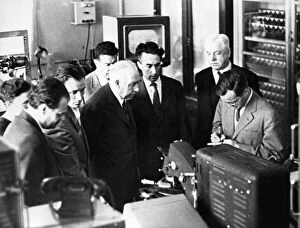
Nils and Aage Bohr in laboratory C016 / 8375
Physicists in a laboratory. Physicists Nils and Aage Bohr being shown an early video-microscope used to study nuclear tracks on photographic emulsions

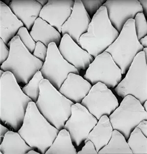
Background imageMicroscope Collection: Scyliorhinus canicula, dogfish

Scyliorhinus canicula, dogfish
Scanning electron microscope (SEM) image of the scales of a dogfish (x 40)

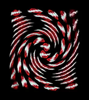
Background imageMicroscope Collection: Dividing cells

Dividing cells. Computer artwork of a spiral of cells during mitosis (nuclear division). During mitosis two daughter nuclei are formed from one parent nucleus

Background imageMicroscope Collection: Nikolai Dubinin, Russian geneticist

Nikolai Dubinin, Russian geneticist
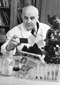
Nikolai Petrovich Dubinin (1907-1998), Russian geneticist, examining microscope slides in a laboratory

Background imageMicroscope Collection: Intestinal microvilli, TEM

Intestinal microvilli, TEM
Intestinal microvilli, coloured transmission electron micrograph (TEM)

Background imageMicroscope Collection: Influenza virus particles, TEM

Influenza virus particles, TEM
Influenza virus particles, coloured transmission electron micrograph (TEM). This virus is the cause of the respiratory disease influenza (flu) in humans and other animals

Background imageMicroscope Collection: Hepatitis C viruses, TEM

Hepatitis C viruses, TEM
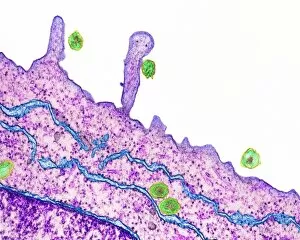
Hepatitis C viruses. Coloured transmission electron micrograph (TEM) of hepatitis C virus particles (green) infecting cultured liver cells (purple)

Background imageMicroscope Collection: E. coli bacterium, TEM

E. coli bacterium, TEM
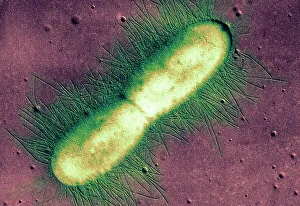
E. coli bacterium. Coloured transmission electron micrograph (TEM) of an Escherichia coli bacterium in the early stages of binary fission, the process by which the bacterium divides

Background imageMicroscope Collection: Manufacture of circuit boards

Manufacture of circuit boards
A complicated multi-component electronic device for manufacturing circuit boards

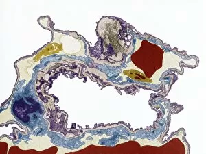
Background imageMicroscope Collection: Lung tissue, TEM

Lung tissue, TEM
Lung tissue, coloured transmission electron micrograph (TEM). A single alveolus is at center. At top right is a pulmonary capillary containing a red blood cell (red) and a platelet (yellow)

Background imageMicroscope Collection: investigating rinderpest at Kimberley, South Africa: drawing, 1897

investigating rinderpest at Kimberley, South Africa: drawing, 1897
ROBERT KOCH (1843-1910) investigating rinderpest at Kimberley, South Africa: drawing, 1897

Background imageMicroscope Collection: Rosenhain optical microscope

Rosenhain optical microscope. The viewing lens (eyepiece, upper right), focusing wheel (lower right) and objective lens (centre) are seen

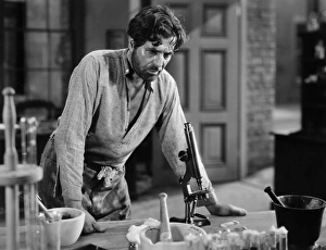
Background imageMicroscope Collection: SILENT STILL: LABORATORIES. Warner Baxter as Dr

SILENT STILL: LABORATORIES. Warner Baxter as Dr. Samuel Mudd, The Prisoner of Shark Island, 1936

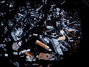
Background imageMicroscope Collection: Microscope image of the Pasamonte eucrite

Microscope image of the Pasamonte eucrite
Microscopic image of the Pasamonte eucrite showing a basaltic texture. Field of view is 2.5mm across

Background imageMicroscope Collection: pine (Pinus spec.), cross section of a few years old stem of a pine, microtome section

pine (Pinus spec.), cross section of a few years old stem of a pine, microtome section

Background imageMicroscope Collection: Mahatma gandhi observing leprosy germs, sevagram ashram, maharashtra, india, asia, 1940

Mahatma gandhi observing leprosy germs, sevagram ashram, maharashtra, india, asia, 1940

Background imageMicroscope Collection: Health. Sample sperm. Donor Sperm Close Concept of Bank Sperm. Sperm on Blue Background

Health. Sample sperm. Donor Sperm Close Concept of Bank Sperm. Sperm on Blue Background Health. Sample sperm. Donor Sperm Close Concept of Bank Sperm. Sperm on Blue Background

Background imageMicroscope Collection: Mayfly magnified, with various moths

Mayfly magnified, with various moths
Mayfly magnified through microscope 1, fanfooted or fan-foot moth, Herminia tarsipennalis 2, mayfly 3, scarce burnished brass, Diachrysia chryson 4, green carpet, Colostygia pectinataria 5

Background imageMicroscope Collection: Interior structure of the broad bean, Vicia faba

Interior structure of the broad bean, Vicia faba. Section through the stem between two leaves viewed under a microscope. Structure interieure d'un Pied de Feve

Background imageMicroscope Collection: Geography symbols seamless pattern

Geography symbols seamless pattern. Equipments for web banners background. Vintage outline sketch for web banners. Doodle style. Education concept Geography symbols seamless pattern

Background imageMicroscope Collection: Microglia cells stained with Rio Hortega's silver carbonate method in the grey matter of the brain

Microglia cells stained with Rio Hortega's silver carbonate method in the grey matter of the brain

Background imageMicroscope Collection: Complex melanocytic nevus. Dermatoscopy of the skin

Complex melanocytic nevus. Dermatoscopy of the skin. Macro magnified 20 times with a dermatoscope microscope. A set of different nevus. Examination by a dermatologist

Background imageMicroscope Collection: Mycobacterium cartoon character design style with board

Mycobacterium cartoon character design style with board Mycobacterium cartoon character design style with board. Vector illustration

Background imageMicroscope Collection: Heliansthus stem micrograph

Heliansthus stem micrograph Light photomicrograph of Helianthus stem cross section seen through microscope

Background imageMicroscope Collection: Antonie van Leeuwenhoek, 1632-1723. Dutch scientist

Antonie van Leeuwenhoek, 1632-1723. Dutch scientist

Background imageMicroscope Collection: Colon cancer tissue section under the microscope 100x

Colon cancer tissue section under the microscope 100x

Background imageMicroscope Collection: Microscope icon, outline style

Microscope icon, outline style Microscope icon in outline style isolated vector illustration

Background imageMicroscope Collection: Roquefort cheese mold (Penicillium roqueforti) spores under the microscope

Roquefort cheese mold (Penicillium roqueforti) spores under the microscope Magnification is about x985 when printed at 10 centimeters wide

Background imageMicroscope Collection: Focus stacked, inverted image of Diatoms on a microscope slide

Focus stacked, inverted image of Diatoms on a microscope slide

Background imageMicroscope Collection: Limestone microfossils in a 0. 03mm-thick slice of fossil-rich limestone viewed at high magnification

Limestone microfossils in a 0. 03mm-thick slice of fossil-rich limestone viewed at high magnification
Limestone microfossils in a 0.03mm-thick slice of fossil-rich limestone viewed at high magnification, image area is 3mm across in real life

Background imageMicroscope Collection: J Lancaster & Son, Colmore Row, Birmingham

J Lancaster & Son, Colmore Row, Birmingham
J Lancaster & Son, Cameras, Photographic and Scientific Equipment, Colmore Row, Birmingham. circa 1900

Background imageMicroscope Collection: Women night workers at Texas Instruments Ltd in Bedford Circa February 1970

Women night workers at Texas Instruments Ltd in Bedford Circa February 1970. Workers seen here grading transistors and integrated circuit boards

Background imageMicroscope Collection: African daisy (Osteospermum jucundum) at approx 10x magnification

African daisy (Osteospermum jucundum) at approx 10x magnification, composite flower showing individual florets full of golden pollen grains, Monmouthshire, Wales, UK, July

Background imageMicroscope Collection: A lacquered brass microscope belonging to the Darwin family, c.1826-30 (mixed media)

A lacquered brass microscope belonging to the Darwin family, c.1826-30 (mixed media)
8736150 A lacquered brass microscope belonging to the Darwin family, c.1826-30 (mixed media) by English School

Background imageMicroscope Collection: A lacquered brass microscope belonging to the Darwin family, c.1826-30 (mixed media)

A lacquered brass microscope belonging to the Darwin family, c.1826-30 (mixed media)
8736140 A lacquered brass microscope belonging to the Darwin family, c.1826-30 (mixed media) by English School

Background imageMicroscope Collection: A lacquered brass microscope belonging to the Darwin family, c.1826-30 (mixed media)

A lacquered brass microscope belonging to the Darwin family, c.1826-30 (mixed media)
8736155 A lacquered brass microscope belonging to the Darwin family, c.1826-30 (mixed media) by English School

Background imageMicroscope Collection: A lacquered brass microscope belonging to the Darwin family, c.1826-30 (mixed media)

A lacquered brass microscope belonging to the Darwin family, c.1826-30 (mixed media)
8736176 A lacquered brass microscope belonging to the Darwin family, c.1826-30 (mixed media) by English School

Background imageMicroscope Collection: Microscope by Adams of London

Microscope by Adams of London
5311766 Microscope by Adams of London; (add.info.: Illustration of a microscope by Adams of London. Dated circa 1788.); Universal History Archive/UIG

Background imageMicroscope Collection: A magnifying glass in a tube, simple microscope, and diminishing lens, 17th century

A magnifying glass in a tube, simple microscope, and diminishing lens, 17th century
5311406 A magnifying glass in a tube, simple microscope, and diminishing lens, 17th century; Universal History Archive/UIG

Background imageMicroscope Collection: Snowflakes seen under the microscope - Snowflake crystals under the microscope

Snowflakes seen under the microscope - Snowflake crystals under the microscope
FLO4576336 Snowflakes seen under the microscope - Snowflake crystals under the microscope

Background imageMicroscope Collection: Threads of Dutch linen 1 and gold 2 magnified under a microscope

Threads of Dutch linen 1 and gold 2 magnified under a microscope
FLO4574760 Threads of Dutch linen 1 and gold 2 magnified under a microscope

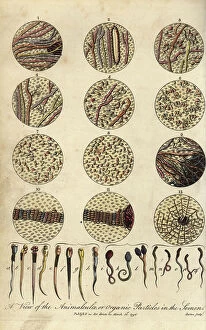
Background imageMicroscope Collection: Microscopic views of human spermatozoa

Microscopic views of human spermatozoa
FLO4714918 Microscopic views of human spermatozoa. View of the animalculae or organic particles in the semen. Handcoloured copperplate engraving by J

Background imageMicroscope Collection: Female American mathematician and astronomer at work, at the Paris Observatory, 1850

Female American mathematician and astronomer at work, at the Paris Observatory, 1850
3478832 Female American mathematician and astronomer at work, at the Paris Observatory, 1850; (add.info.: Miss Klumpke an American mathematician and astronomer at work on the Carte du Ciel)

Background imageMicroscope Collection: Title page of the first volume of Robert Brown's Science for All, 1850

Title page of the first volume of Robert Brown's Science for All, 1850
3331022 Title page of the first volume of Robert Brown's Science for All, 1850; (add.info.: Title page of the first volume of Robert Brown's Science for All')

Background imageMicroscope Collection: Micrographia

Micrographia
2626678 Micrographia; (add.info.: Illustration from Micrographia 1665; by Robert Hooke, 1635-1703)

Background imageMicroscope Collection: Cow tongue under the microscope: veal tongue showing different papillae 1

Cow tongue under the microscope: veal tongue showing different papillae 1
FLO4574832 Cow tongue under the microscope: veal tongue showing different papillae 1, foliate papilla 2, filiform papillae 3, and fungiform papillae 4

Background imageMicroscope Collection: Single microscope mechanisms and parts

Single microscope mechanisms and parts
FLO4593776 Single microscope mechanisms and parts. Copperplate engraving by Wilson Lowry after a drawing by J. Farey Jr

Background imageMicroscope Collection: Projection and enlargement of microscopic reports during the siege of Paris in 1871

Projection and enlargement of microscopic reports during the siege of Paris in 1871
PCT4256951 Projection and enlargement of microscopic reports during the siege of Paris in 1871

Background imageMicroscope Collection: Crystals magnified under a microscope: silver solution and Diana's tree 1

Crystals magnified under a microscope: silver solution and Diana's tree 1
FLO4574143 Crystals magnified under a microscope: silver solution and Diana's tree 1, camphor 2, and niter 3

Background imageMicroscope Collection: Common fly, Musca domestica 1, head of fly 2

Common fly, Musca domestica 1, head of fly 2
FLO4574014 Common fly, Musca domestica 1, head of fly 2, magnified 3, proboscis 4, 5, cornea 6, magnified 7

Background imageMicroscope Collection: Three pillar microscope

Three pillar microscope
5311760 Three pillar microscope; (add.info.: Illustration of a Three pillar microscope of the type produced by Edmund Culpeper (1660-1738) circa 1730.); Universal History Archive/UIG

Background imageMicroscope Collection: Micrographia

Micrographia
2626676 Micrographia; (add.info.: Illustration from Micrographia 1665; by Robert Hooke, 1635-1703)

Background imageMicroscope Collection: Comparison with microscope between the regularite of the yarn of the yarn of the araignee

Comparison with microscope between the regularite of the yarn of the yarn of the araignee
FLO4576287 Comparison with microscope between the regularite of the yarn of the yarn of the araignee and the inegal weaving of the lace yarns - Observations under the microscope

Background imageMicroscope Collection: France, Rhone-Alpes, Rhone (69)

France, Rhone-Alpes, Rhone (69)
LUX4811904 France, Rhone-Alpes, Rhone (69), Lyon: the interior of a laboratory with a biologist posing in working clothes with a rabbit of experimentation, 1900 - Lot Z; (add.info.: France)

Background imageMicroscope Collection: A lacquered brass microscope belonging to the Darwin family, c.1826-30 (mixed media)

A lacquered brass microscope belonging to the Darwin family, c.1826-30 (mixed media)
8736188 A lacquered brass microscope belonging to the Darwin family, c.1826-30 (mixed media) by English School

Background imageMicroscope Collection: A lacquered brass microscope belonging to the Darwin family, c.1826-30 (mixed media)

A lacquered brass microscope belonging to the Darwin family, c.1826-30 (mixed media)
8736162 A lacquered brass microscope belonging to the Darwin family, c.1826-30 (mixed media) by English School

Background imageMicroscope Collection: How man learns from the human eye (litho)

How man learns from the human eye (litho)
8676259 How man learns from the human eye (litho) by English School, (20th century); Private Collection; (add.info.: How man learns from the human eye. Inventions using lenses)

Background imageMicroscope Collection: Monkey examining food through a microscope (colour litho)

Monkey examining food through a microscope (colour litho)
8656327 Monkey examining food through a microscope (colour litho) by English School

Background imageMicroscope Collection: Compound microscope

Compound microscope
5311762 Compound microscope; (add.info.: Illustration depicting a compound microscope by Adams of London. Dated circa 1788.); Universal History Archive/UIG

Background imageMicroscope Collection: A Zeiss of Jena microscope with movable alternative objectives

A Zeiss of Jena microscope with movable alternative objectives
5311759 A Zeiss of Jena microscope with movable alternative objectives; (add.info.: Engraving depicting a Zeiss of Jena microscope with movable alternative objectives)

Background imageMicroscope Collection: Illustration showing the examination of a live frog under a microscope. German, 1895

Illustration showing the examination of a live frog under a microscope. German, 1895
5311517 Illustration showing the examination of a live frog under a microscope. German, 1895; (add.info.: Date: 1895); Universal History Archive/UIG

Background imageMicroscope Collection: Magnified image under a microscope of the fibres of a sponge 1

Magnified image under a microscope of the fibres of a sponge 1
FLO4576246 Magnified image under a microscope of the fibres of a sponge 1, and two-tone taffeta fabric, with yellow weft and purple warp 2

Background imageMicroscope Collection: France, Rhone-Alpes, Rhone (69)

France, Rhone-Alpes, Rhone (69)
LUX4811888 France, Rhone-Alpes, Rhone (69), Lyon: The interior of a laboratory with a biologist posing in a work suit, 1900 - Lot Z; (add.info.: France, Rhone-Alpes, Rhone (69)

Background imageMicroscope Collection: Compound microscope mechanisms and parts including Adams

Compound microscope mechanisms and parts including Adams
FLO4593789 Compound microscope mechanisms and parts including Adams and Martin's Copperplate engraving by Wilson Lowry after a drawing by J


All products are expertly crafted, using premium materials, tailored to your specifications and promptly shipped

"Unveiling the Unseen: Exploring the Microscopic World" Step into a realm of hidden wonders, where scientists like Rosalind Franklin and Santiago Ramon y Cajal have unraveled the mysteries through their microscopic lens. From Liver Fluke to Norovirus particles, these pioneers have paved the way for groundbreaking discoveries. Witness the intricate beauty of Snail teeth and Fat cells as they come alive under the microscope's gaze. Marvel at Lubbock's Graphic 84, a visual masterpiece capturing the essence of this microcosmic universe. Delve deeper into this microscopic world and encounter Plasmodium sp. , a malarial parasite that has plagued humanity for centuries. Explore E. Coli bacterium in all its glory, revealing both its potential dangers and scientific significance. Through microscope slide preparation techniques, we gain insight into complex organisms like never before. Discover how Fleming used his photographic skills to document crucial moments in microbiology history. The microscope acts as our window into an unseen dimension - it allows us to explore realms beyond our naked eye's reach, and is through this powerful tool that we continue to unravel nature's secrets one magnified image at a time. So join us on this journey "Under the Microscope, " where every slide holds a story waiting to be told – stories that shape our understanding of life itself.

© Copyright Media Storehouse, All Rights Reserved 2003 - 2024
Facebook     Pinterest     100% Payment Secure     Reviews IO