Fraud Blocker Skip to main content

Microscope Collection (page 15)

"Unveiling the Unseen: Exploring the Microscopic World" Step into a realm of hidden wonders

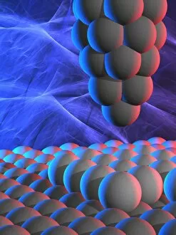
Background imageMicroscope Collection: Scanning tunnelling microscope, artwork F005 / 0813

Scanning tunnelling microscope, artwork F005 / 0813
Scanning tunnelling microscope (STM), computer artwork. The STM uses a fine needle with an electric potential running through it to scan the surface of an object
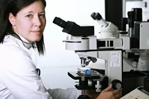
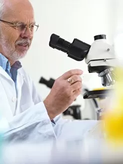
Background imageMicroscope Collection: Biological research F005 / 0936

Biological research F005 / 0936
MODEL RELEASED. Biological research. Researcher using a light microscope

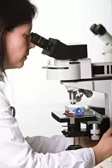
Background imageMicroscope Collection: Biological research F005 / 0937

Biological research F005 / 0937
MODEL RELEASED. Biological research. Researcher using a light microscope

Background imageMicroscope Collection: H1N1 swine flu virus, TEM C018 / 8602

H1N1 swine flu virus, TEM C018 / 8602
H1N1 swine flu virus. Coloured transmission electron micrograph (TEM) of swine influenza (flu) virus particles (virions, yellow)

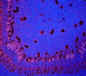
Background imageMicroscope Collection: Cotton stem, confocal micrograph C014 / 4636

Cotton stem, confocal micrograph C014 / 4636
Cotton stem. Laser scanning confocal micrograph (LSCM) of a section through the stem of a cotton (Gossypium sp.) plant

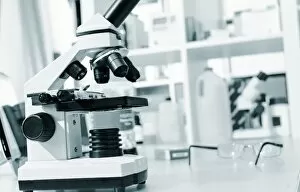
Background imageMicroscope Collection: Light microscope C014 / 1407

Light microscope C014 / 1407
Light microscope, seen in silhouetted profile. This microscope has a binocular eyepiece (an eyepiece for each eye). The sample is placed on a glass slide on the mount below centre

Background imageMicroscope Collection: Microscopic Vegetable Objects C017 / 3506

Microscopic Vegetable Objects C017 / 3506
Plate from 18th century encyclopedia showing illustrations of microscopical objects. Shown are various seeds of plants including; apple, clematis, columbine, cabbage, cucumber, jasmine, pear, poppy

Background imageMicroscope Collection: Biological research F007 / 0378

Biological research F007 / 0378
Biological research

Background imageMicroscope Collection: Light microscope F007 / 0356

Light microscope F007 / 0356
Light microscope

Background imageMicroscope Collection: Microbiology research F007 / 0304

Microbiology research F007 / 0304
MODEL RELEASED. Microbiology research

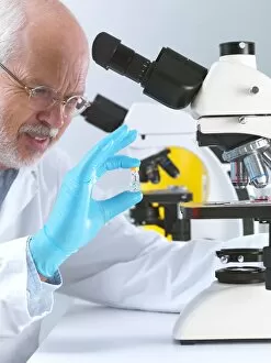
Background imageMicroscope Collection: Scientist using a light microscope F006 / 9023

Scientist using a light microscope F006 / 9023
MODEL RELEASED. Scientist using a light microscope

Background imageMicroscope Collection: Biological research F006 / 9024

Biological research F006 / 9024
MODEL RELEASED. Biological research

Background imageMicroscope Collection: Biological research F006 / 9025

Biological research F006 / 9025
MODEL RELEASED. Biological research

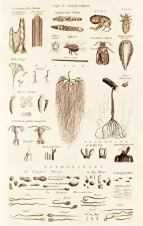
Background imageMicroscope Collection: Microscopical Objects C017 / 3449

Microscopical Objects C017 / 3449
Plate from 18th century encyclopedia showing illustrations of microscopical objects including small animals and parts of animals, microscopic or minute organisms, such as rotifers

Background imageMicroscope Collection: Light microscope F006 / 8553

Light microscope F006 / 8553
Light microscope

Background imageMicroscope Collection: Light microscope F006 / 8554

Light microscope F006 / 8554
Light microscope

Background imageMicroscope Collection: Light microscope F006 / 8551

Light microscope F006 / 8551
Light microscope

Background imageMicroscope Collection: Robert Hooke, English natural philosopher C017 / 7121

Robert Hooke, English natural philosopher C017 / 7121
Robert Hooke (1635-1703), English natural philosopher. Hooke made numerous advances in fields as diverse as astronomy, physics and microbiology, and his name is remembered in Hookes Law

Background imageMicroscope Collection: Light microscope F006 / 9033

Light microscope F006 / 9033
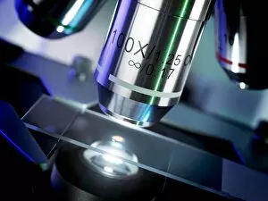
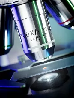
Light microscope. Close up of a slide on a microscope stage

Background imageMicroscope Collection: Light microscope F006 / 9034

Light microscope F006 / 9034
Light microscope. Close up of a slide on a microscope stage

Background imageMicroscope Collection: Light microscope F006 / 9032

Light microscope F006 / 9032
Light microscope. Close up of a slide on a microscope stage

Background imageMicroscope Collection: Haematology lab F006 / 9031

Haematology lab F006 / 9031
MODEL RELEASED. Haematology lab

Background imageMicroscope Collection: Microbiology research F006 / 9028

Microbiology research F006 / 9028
MODEL RELEASED. Microbiology research

Background imageMicroscope Collection: Microbiology research F006 / 9027

Microbiology research F006 / 9027
MODEL RELEASED. Microbiology research

Background imageMicroscope Collection: Scientist using a light microscope F006 / 9029

Scientist using a light microscope F006 / 9029
MODEL RELEASED. Scientist using a light microscope

Background imageMicroscope Collection: Haematology lab F006 / 9030

Haematology lab F006 / 9030
MODEL RELEASED. Haematology lab

Background imageMicroscope Collection: Biological research F006 / 9026

Biological research F006 / 9026
MODEL RELEASED. Biological research

Background imageMicroscope Collection: High-contrast direct DNA image, TEM

High-contrast direct DNA image, TEM
High-contrast direct DNA image. Coloured transmission electron micrograph (TEM) of the first high-contrast direct image of a bundle (fibre) of strands of DNA (deoxyribonucleic acid)

Background imageMicroscope Collection: Laser microscope experiment C016 / 6434

Laser microscope experiment C016 / 6434
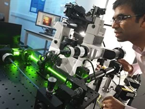
Laser microscope experiment. Researcher working in a laboratory on an experiment with lasers (green light) and a microscope. The microscope being used here is a type known as an inverted microscope

Background imageMicroscope Collection: Laser microscope experiment C016 / 6430

Laser microscope experiment C016 / 6430
Laser microscope experiment. Researcher working in a laboratory on an experiment with lasers (green light) and a microscope. The microscope being used here is a type known as an inverted microscope

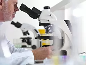
Background imageMicroscope Collection: Light microscopy C016 / 9700

Light microscopy C016 / 9700
Light microscopy. Light microscope being used to magnify a blood sample

Background imageMicroscope Collection: Light microscopy C016 / 9703

Light microscopy C016 / 9703
Light microscopy. Light microscope being used to magnify a blood sample

Background imageMicroscope Collection: Roundworm germ cells, light micrograph C016 / 9538

Roundworm germ cells, light micrograph C016 / 9538
Roundworm germ cells. Light micrograph of germ cells from a roundworm (Ascaris sp.), undergoing mitosis (nuclear division)

Background imageMicroscope Collection: Light microscopy C016 / 9702

Light microscopy C016 / 9702
Light microscopy. Light microscope being used to magnify a blood sample

Background imageMicroscope Collection: Light microscopy C016 / 9701

Light microscopy C016 / 9701
Light microscopy. Light microscope being used to magnify a blood sample

Background imageMicroscope Collection: Schmallenberg virus particles, TEM

Schmallenberg virus particles, TEM
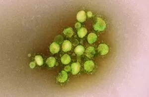
Schmallenberg virus. Transmission electron micrograph (TEM) of virus particles (virions, yellow) of the Schmallenberg virus

Background imageMicroscope Collection: Activated plasma cell, TEM

Activated plasma cell, TEM
Activated plasma cell, coloured transmission electron micrograph (TEM). Plasma cells are derived from stimulated B lymphocytes (white blood cells)

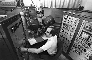
Background imageMicroscope Collection: Controls of early electron microscope

Controls of early electron microscope
Electron microscope. View of a technician operating the controls of an early model of electron microscope. The cabinets either side of the frame are computers and control equipment

Background imageMicroscope Collection: MERS coronavirus, TEM C015 / 1772

MERS coronavirus, TEM C015 / 1772
MERS coronavirus, coloured transmission electron micrograph (TEM). This virus (originally novel coronavirus 2012) has been named Middle East respiratory syndrome (MERS) coronavirus

Background imageMicroscope Collection: MERS coronavirus, TEM C015 / 1774

MERS coronavirus, TEM C015 / 1774
MERS coronavirus, coloured transmission electron micrograph (TEM). This virus (originally novel coronavirus 2012) has been named Middle East respiratory syndrome (MERS) coronavirus

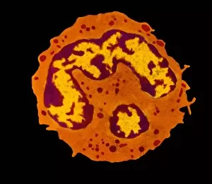
Background imageMicroscope Collection: Granulocyte white blood cell, TEM

Granulocyte white blood cell, TEM
Granulocyte white blood cell. Coloured transmission electron micrograph (TEM) of a neutrophil granulocyte, a type of white blood cell

Background imageMicroscope Collection: MERS coronavirus, TEM C015 / 1776

MERS coronavirus, TEM C015 / 1776
MERS coronavirus, coloured transmission electron micrograph (TEM). This virus (originally novel coronavirus 2012) has been named Middle East respiratory syndrome (MERS) coronavirus

Background imageMicroscope Collection: MERS coronavirus, TEM C015 / 1773

MERS coronavirus, TEM C015 / 1773
MERS coronavirus, coloured transmission electron micrograph (TEM). This virus (originally novel coronavirus 2012) has been named Middle East respiratory syndrome (MERS) coronavirus

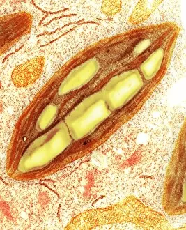
Background imageMicroscope Collection: Chloroplast, TEM C016 / 6297

Chloroplast, TEM C016 / 6297
Chloroplast. Coloured transmission electron micrograph (TEM) of chloroplast from the moss Physcomitrella patens. Chloroplasts are the sites of photosynthesis

Background imageMicroscope Collection: Chloroplast, TEM C016 / 6298

Chloroplast, TEM C016 / 6298
Chloroplast. Coloured transmission electron micrograph (TEM) of chloroplast from the moss Physcomitrella patens. Chloroplasts are the sites of photosynthesis

Background imageMicroscope Collection: Measles virus particle, TEM C015 / 7160

Measles virus particle, TEM C015 / 7160
Measles virus particle, coloured transmission electron micrograph (TEM). This virus, from the Morbillivirus group of viruses, consists of an RNA (ribonucleic acid)

Background imageMicroscope Collection: Measles virus particle, TEM C015 / 7161

Measles virus particle, TEM C015 / 7161
Measles virus particle, coloured transmission electron micrograph (TEM). This virus, from the Morbillivirus group of viruses, consists of an RNA (ribonucleic acid)

Background imageMicroscope Collection: MERS coronavirus, TEM C015 / 7155

MERS coronavirus, TEM C015 / 7155
MERS coronavirus. Coloured transmission electron micrograph (TEM) of MERS coronavirus particles (orange) budding from a host cell (brown)


All Professionally Made to Order for Quick Shipping

"Unveiling the Unseen: Exploring the Microscopic World" Step into a realm of hidden wonders, where scientists like Rosalind Franklin and Santiago Ramon y Cajal have unraveled the mysteries through their microscopic lens. From Liver Fluke to Norovirus particles, these pioneers have paved the way for groundbreaking discoveries. Witness the intricate beauty of Snail teeth and Fat cells as they come alive under the microscope's gaze. Marvel at Lubbock's Graphic 84, a visual masterpiece capturing the essence of this microcosmic universe. Delve deeper into this microscopic world and encounter Plasmodium sp. , a malarial parasite that has plagued humanity for centuries. Explore E. Coli bacterium in all its glory, revealing both its potential dangers and scientific significance. Through microscope slide preparation techniques, we gain insight into complex organisms like never before. Discover how Fleming used his photographic skills to document crucial moments in microbiology history. The microscope acts as our window into an unseen dimension - it allows us to explore realms beyond our naked eye's reach, and is through this powerful tool that we continue to unravel nature's secrets one magnified image at a time. So join us on this journey "Under the Microscope, " where every slide holds a story waiting to be told – stories that shape our understanding of life itself.