Fraud Blocker Skip to main content

Microscopy Collection

Microscopy, the art of exploring the hidden world beyond our naked eyes, unveils breathtaking wonders that captivate both scientists and artists alike

Background imageMicroscopy Collection: Bee anatomy, historical artwork

Bee anatomy, historical artwork
Bees anatomy. 17th Century artwork by the Italian microscopist Francesco Stelluti, showing the anatomy of bees as seen by him under a microscope

Background imageMicroscopy Collection: EDTA crystals, light micrograph

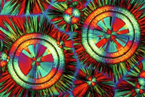
EDTA crystals, light micrograph
EDTA crystals. Polarised light micrograph of a section through ethylenediaminetetraacetic acid (EDTA) crystals. EDTA is used primarily for its effectiveness at binding metal ions (chelating)

Background imageMicroscopy Collection: Illustration of a Flea C017 / 3435

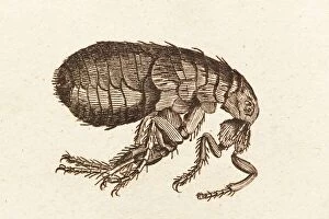
Illustration of a Flea C017 / 3435
Plate from 18th century encyclopedia showing an illustration of a flea. Fleas are the insects forming the order Siphonaptera and are a vectors for various diseases

Background imageMicroscopy Collection: Snowflake research, 18th century

Snowflake research, 18th century

Background imageMicroscopy Collection: Pine pollen grains, light micrograph

Pine pollen grains, light micrograph
Pine pollen grains. Light micrograph of a section through winged pollen grains (microspores) from a pine (Pinus sp.) tree

Background imageMicroscopy Collection: Pine stem, light micrograph

Pine stem, light micrograph
Pine stem. Light micrograph of a section through the stem of a pine (Pinus sp.) tree, showing xylem tissue. The xylem is made up of tracheid cells (light pink)

Background imageMicroscopy Collection: Lime tree stem, light micrograph

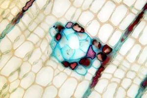
Lime tree stem, light micrograph
Lime tree stem. Light micrograph of a section through the stem of a lime tree (Tilia europaea). The outer epidermis has been shed and replaced by a layer of cork (dark red)

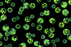
Background imageMicroscopy Collection: Water fleas (Daphnia sp. ) and a green algae (Volvox aureus) in water from a garden pond

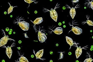
Water fleas (Daphnia sp. ) and a green algae (Volvox aureus) in water from a garden pond
Water fleas (Daphnia sp.) and a green algae (Volvox aureus) in water from a garden pond. Derbyshire, UK. September. Digital composite

Background imageMicroscopy Collection: Chlorophyte or green alga (Volvox aureus) in pond water. UK. The larger spheres are approximately 0

Chlorophyte or green alga (Volvox aureus) in pond water. UK. The larger spheres are approximately 0.5mm in diameter. Digital composite image

Background imageMicroscopy Collection: Portrait of a Common Wasp (Vespula vulgaris). UK. Focus stacked image (dead specimen)

Portrait of a Common Wasp (Vespula vulgaris). UK. Focus stacked image (dead specimen)

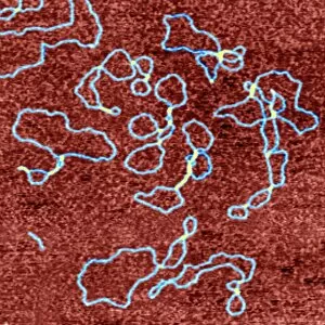
Background imageMicroscopy Collection: Anti-cancer drug binding to DNA, AFM

Anti-cancer drug binding to DNA, AFM
Drug-DNA complexes. Coloured atomic force micrograph (AFM) of plasmids (blue) of DNA (deoxyribonucleic acid) bound to the anti-cancer drug ditercalinium

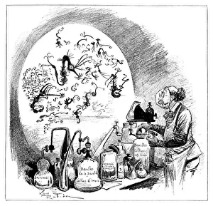
Background imageMicroscopy Collection: Microbiology caricature, 19th century

Microbiology caricature, 19th century
Microbiology caricature. The researcher is handling jars labelled as bacterial cultures, while the magnified view at left shows a caricatured appearance of microbes as seen under a microscope

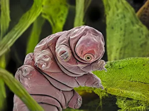
Background imageMicroscopy Collection: False-coloured SEM (Scanning Electron Micrograph) of a Tardigrade (Tardigrada) amongst moss

False-coloured SEM (Scanning Electron Micrograph) of a Tardigrade (Tardigrada) amongst moss, from Peak District National Park, Derbyshire, UK. This tardigrade is approximately 0.1 millimetres long

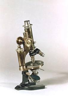
Background imageMicroscopy Collection: Rosenhain optical microscope

Rosenhain optical microscope. The viewing lens (eyepiece, upper right), focusing wheel (lower right) and objective lens (centre) are seen

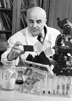
Background imageMicroscopy Collection: Nikolai Dubinin, Russian geneticist

Nikolai Dubinin, Russian geneticist
Nikolai Petrovich Dubinin (1907-1998), Russian geneticist, examining microscope slides in a laboratory. Dubinin was one of the founding members of the Cytology

Background imageMicroscopy Collection: Micrographia

Micrographia
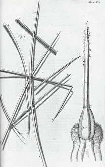
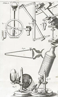
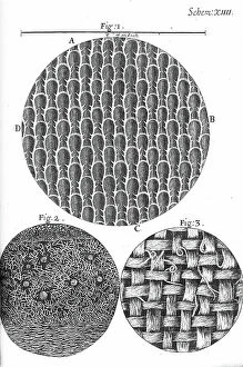
2626678 Micrographia; (add.info.: Illustration from 'Micrographia' 1665; by Robert Hooke, 1635-1703. Hooke's observations through various lenses)

Background imageMicroscopy Collection: Micrographia

Micrographia
2626676 Micrographia; (add.info.: Illustration from 'Micrographia' 1665; by Robert Hooke, 1635-1703. Hooke's observations through various lenses)

Background imageMicroscopy Collection: Illustration showing the examination of a live frog under a microscope. German, 1895

Illustration showing the examination of a live frog under a microscope. German, 1895
5311517 Illustration showing the examination of a live frog under a microscope. German, 1895; (add.info.: Date: 1895); Universal History Archive/UIG.

Background imageMicroscopy Collection: Micrographia

Micrographia
2626674 Micrographia; (add.info.: Illustration from 'Micrographia' 1665; by Robert Hooke, 1635-1703. Hooke's observations through various lenses)

Background imageMicroscopy Collection: Micrographia

Micrographia
2626680 Micrographia; (add.info.: Illustration from 'Micrographia' 1665; by Robert Hooke, 1635-1703. Hooke's observations through various lenses)

Background imageMicroscopy Collection: Micrographia

Micrographia
2626677 Micrographia; (add.info.: Illustration from 'Micrographia' 1665; by Robert Hooke, 1635-1703. Hooke's observations through various lenses)

Background imageMicroscopy Collection: Micrographia

Micrographia
2626673 Micrographia; (add.info.: Illustration from 'Micrographia' 1665; by Robert Hooke, 1635-1703. Hooke's observations through various lenses)

Background imageMicroscopy Collection: Micrographia

Micrographia
2626671 Micrographia; (add.info.: Illustration from 'Micrographia' 1665; by Robert Hooke, 1635-1703. Hooke's observations through various lenses)

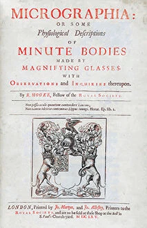
Background imageMicroscopy Collection: Title page Illustration from 'Micrographia'

Title page Illustration from 'Micrographia'
2626681 Title page Illustration from 'Micrographia'; (add.info.: Title page with Illustration from 'Micrographia' 1665; by Robert Hooke, 1635-1703)

Background imageMicroscopy Collection: Micrographia

Micrographia
2626672 Micrographia; (add.info.: Illustration from 'Micrographia' 1665; by Robert Hooke, 1635-1703. Hooke's observations through various lenses)

Background imageMicroscopy Collection: Micrographia

Micrographia
2626679 Micrographia; (add.info.: Illustration from 'Micrographia' 1665; by Robert Hooke, 1635-1703. Hooke's observations through various lenses)

Background imageMicroscopy Collection: Micrographia

Micrographia
2626675 Micrographia; (add.info.: Illustration from 'Micrographia' 1665; by Robert Hooke, 1635-1703. Hooke's observations through various lenses)

Background imageMicroscopy Collection: The Late Mr. Robert Brown, Keeper of Botany in the British Museum... 1858. Creator: Unknown

The Late Mr. Robert Brown, Keeper of Botany in the British Museum... 1858. Creator: Unknown
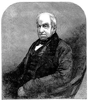
The Late Mr. Robert Brown, Keeper of Botany in the British Museum, from a photograph by Maull and Polyblank, 1858. Engraving from a photograph by Maull and Polyblank

Background imageMicroscopy Collection: Cuttlefish and Gorita, 1785. Creator: Jan Brandes

Cuttlefish and Gorita, 1785. Creator: Jan Brandes
Cuttlefish and Gorita, 1785. Sepia loligo'. Two colour drawings, and a sketch of an octopus-like creature. With inscriptions. Part of Jan Brandes sketchbook, dl. 1 (1808), p. 238

Background imageMicroscopy Collection: The Testimonial presented to Dr. Hassall, on Thursday, 1856. Creator: Unknown

The Testimonial presented to Dr. Hassall, on Thursday, 1856. Creator: Unknown
The Testimonial presented to Dr. Hassall, on Thursday, 1856. Gift to British chemist and microscopist Arthur Hill Hassall

Background imageMicroscopy Collection: Focus stacked, inverted image of Diatoms on a microscope slide

Focus stacked, inverted image of Diatoms on a microscope slide. Diatoms are single-celled algae which produce approximately 25% of the oxygen we breathe as well as being responsible for approximately

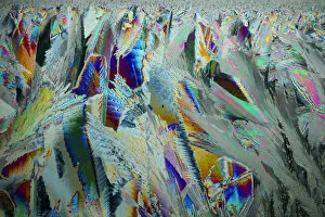
Background imageMicroscopy Collection: Sheet ice crystals viewed by polarised light

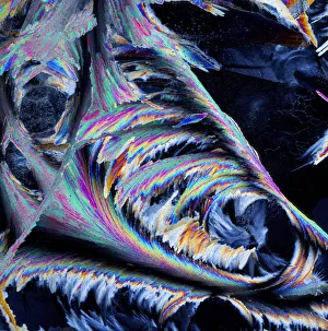
Sheet ice crystals viewed by polarised light

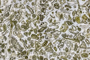
Background imageMicroscopy Collection: Limestone microfossils in a 0. 03mm-thick slice of fossil-rich limestone viewed at high

Limestone microfossils in a 0. 03mm-thick slice of fossil-rich limestone viewed at high magnification
Limestone microfossils in a 0.03mm-thick slice of fossil-rich limestone viewed at high magnification, image area is 3mm across in real life

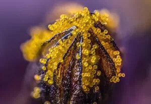
Background imageMicroscopy Collection: African daisy (Osteospermum jucundum) at approx 10x magnification

African daisy (Osteospermum jucundum) at approx 10x magnification, composite flower showing individual florets full of golden pollen grains, Monmouthshire, Wales, UK, July

Background imageMicroscopy Collection: Plankton at night, Palau, Philippine Sea

Plankton at night, Palau, Philippine Sea

Background imageMicroscopy Collection: Ice crystals viewed by polarised light

Ice crystals viewed by polarised light

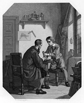
Background imageMicroscopy Collection: Hans Jansen and his son Sacharias, c1870. Artist: H Sluyter

Hans Jansen and his son Sacharias, c1870. Artist: H Sluyter
Hans Jansen and his son Sacharias, c1870. Some historians credit Sacharias Jansen, a Middelburg spectacle maker, with the invention of the telescope and the microscope

Background imageMicroscopy Collection: Microscope made by C Reichert, Vienna, 1895. Artist: C Reichert

Microscope made by C Reichert, Vienna, 1895. Artist: C Reichert
Microscope made by C Reichert, Vienna, 1895. The microscope featured objectives of different powers which could be turned into viewing position as required

Background imageMicroscopy Collection: Colony of Haemophilus influenzae, 1906 (litho)

Colony of Haemophilus influenzae, 1906 (litho)
3501914 Colony of Haemophilus influenzae, 1906 (litho) by German School, (20th century); Private Collection; (add.info.: Bacteria constitute a large domain of prokaryotic microorganisms)

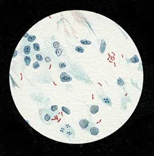
Background imageMicroscopy Collection: Colony of Mycobacterium leprae, 1906 (litho)

Colony of Mycobacterium leprae, 1906 (litho)
3501912 Colony of Mycobacterium leprae, 1906 (litho) by German School, (20th century); Private Collection; (add.info.: Bacteria constitute a large domain of prokaryotic microorganisms)

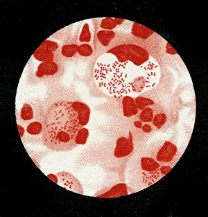
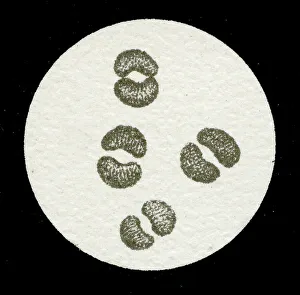
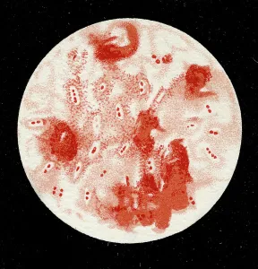
Background imageMicroscopy Collection: Colony of Micrococcus Gonorrhoea, 1906 (litho)

Colony of Micrococcus Gonorrhoea, 1906 (litho)
3501903 Colony of Micrococcus Gonorrhoea, 1906 (litho) by German School, (20th century); Private Collection; (add.info.: Bacteria constitute a large domain of prokaryotic microorganisms)

Background imageMicroscopy Collection: Colony of Streptococcus pneumoniae with and without bubble capsule, 1906 (litho)

Colony of Streptococcus pneumoniae with and without bubble capsule, 1906 (litho)
3501899 Colony of Streptococcus pneumoniae with and without bubble capsule, 1906 (litho) by German School, (20th century); Private Collection; (add.info)

Background imageMicroscopy Collection: Colony of Streptococcus Pneumoniae, 1906 (litho)

Colony of Streptococcus Pneumoniae, 1906 (litho)
3501898 Colony of Streptococcus Pneumoniae, 1906 (litho) by German School, (20th century); Private Collection; (add.info.: Bacteria constitute a large domain of prokaryotic microorganisms)

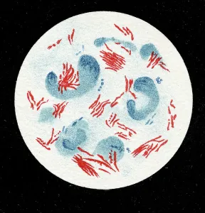
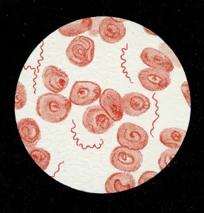
Background imageMicroscopy Collection: Spirillum of the Lyme disease causing bacteria, Spirochaetes Borrelia Recurrentis

Spirillum of the Lyme disease causing bacteria, Spirochaetes Borrelia Recurrentis, in blood sample, 1906 (litho)
3501919 Spirillum of the Lyme disease causing bacteria, Spirochaetes Borrelia Recurrentis, in blood sample, 1906 (litho) by German School

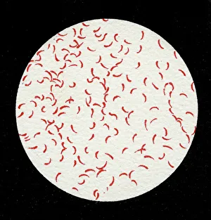
Background imageMicroscopy Collection: Colony of Vibrio cholerae, 1906 (litho)

Colony of Vibrio cholerae, 1906 (litho)
3501918 Colony of Vibrio cholerae, 1906 (litho) by German School, (20th century); Private Collection; (add.info.: Bacteria constitute a large domain of prokaryotic microorganisms)

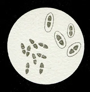
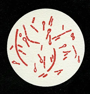
Background imageMicroscopy Collection: Colony of Clostridium tetani with spores, 1906 (litho)

Colony of Clostridium tetani with spores, 1906 (litho)
3501913 Colony of Clostridium tetani with spores, 1906 (litho) by German School, (20th century); Private Collection; (add.info.: Bacteria constitute a large domain of prokaryotic microorganisms)

Background imageMicroscopy Collection: Colony of Mycobacterium tuberculosis, 1906 (litho)

Colony of Mycobacterium tuberculosis, 1906 (litho)
3501910 Colony of Mycobacterium tuberculosis, 1906 (litho) by German School, (20th century); Private Collection; (add.info.: Bacteria constitute a large domain of prokaryotic microorganisms)

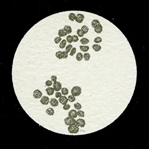
Background imageMicroscopy Collection: A colony of Staphylococcus pyogenes after cell division, 1906 (litho)

A colony of Staphylococcus pyogenes after cell division, 1906 (litho)
3501897 A colony of Staphylococcus pyogenes after cell division, 1906 (litho) by German School, (20th century); Private Collection; (add.info)


All Professionally Made to Order for Quick Shipping

Microscopy, the art of exploring the hidden world beyond our naked eyes, unveils breathtaking wonders that captivate both scientists and artists alike. From intricate bee anatomy to historical artwork, each specimen under the microscope tells a unique story. In the depths of a laboratory, EDTA crystals shimmer like tiny diamonds as they reveal their molecular secrets through a mesmerizing light micrograph. Meanwhile, an illustration of a Flea C017/3435 transports us back in time, reminding us of the meticulous observations made by early pioneers. Snowflake research in the 18th century takes center stage as delicate ice crystals dance before our eyes. Pine pollen grains and pine stems come alive under the lens, showcasing their intricate structures with stunning clarity. The lime tree stem follows suit, revealing its hidden beauty through another enchanting light micrograph. Anton van Leeuwenhoek's groundbreaking discoveries come to life as we gaze upon his meticulously drawn animalcules from c1795. These microscopic organisms remind us of how far we have come in unraveling nature's mysteries. Water fleas and green algae form a delicate ecosystem within garden pond water—a harmonious dance captured for eternity by microscopy's watchful eye. Chlorophyte or green alga Volvox aureus floats gracefully in UK pond water—its larger spheres measuring just about 0—an awe-inspiring sight that reminds us of nature's infinite diversity. An abstract close-up reveals butterfly wing scales' vibrant colors and intricate patterns—a testament to evolution's artistic touch on even the tiniest creatures. And finally, false-colored SEM showcases a Tardigrade amongst moss from Peak District National Park—a minuscule creature measuring only approximately 0. 1 millimeters long but holding immense fascination for researchers worldwide.