
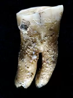
Molar Collection
"Molar: The Mighty Tooth in Leonardo da Vinci's Skull Anatomy" Discover the intricate world of teeth through the eyes of Leonardo da Vinci


All Professionally Made to Order for Quick Shipping
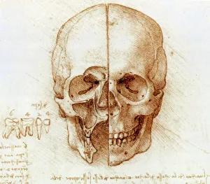
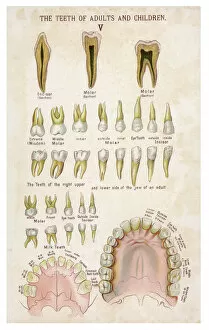
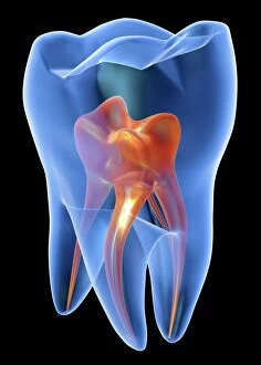
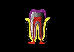
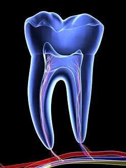
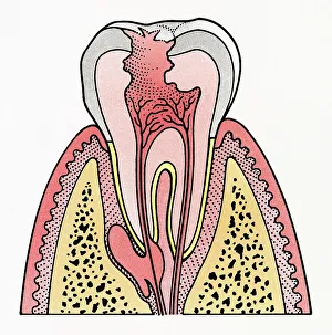
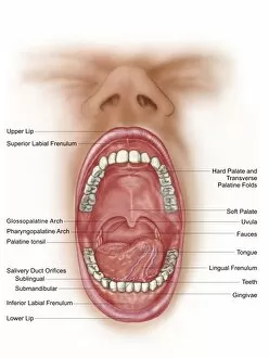
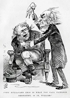
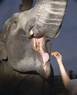
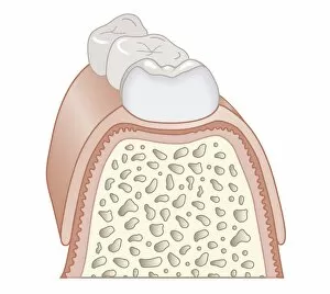
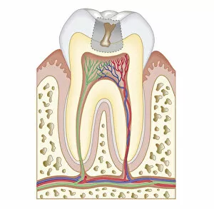
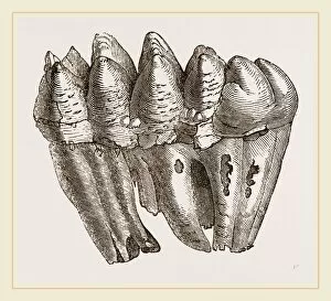
"Molar: The Mighty Tooth in Leonardo da Vinci's Skull Anatomy" Discover the intricate world of teeth through the eyes of Leonardo da Vinci, as he delves into the fascinating realm anatomy. In his iconic artwork, da Vinci meticulously portrays the teeth of both adults and children, shedding light on their unique characteristics. One cannot overlook the prominence given to the mighty molar tooth. Positioned strategically at the back of our mouths, these molars play a crucial role in chewing and grinding food for digestion. Da Vinci's attention to detail captures every aspect of this remarkable tooth with precision. As we explore human tooth anatomy through artistry, we are captivated by wide-open mouths revealing not only molars but also an array of other dental structures such as teeth, tongue, palate, and uvula, and is within this complex mouth cavity that countless stories unfold – from laughter to speech and everything in between. Even cartoons have found humor in dentistry. John Bull finds himself sitting nervously in William Gladstone's dentist chair; a comical representation reminding us that even historical figures had their fair share of dental woes. The Teeth of Adults and Children come alive through vibrant lithographs showcasing their diversity. From erupting milk teeth to permanent ones taking shape - each stage tells a tale about growth and development within our oral cavities. Page after page from The Pictorial Museum of Animated Nature takes us on an enchanting journey deep into nature's wonders. Amongst its engravings lies a captivating depiction illustrating human mouth open - unveiling an amalgamation of teeth, gums, and tongue working harmoniously together. To truly understand our dental structure better, labels guide us through various types of human teeth - incisors for cutting or canines for tearing apart food items before reaching those powerful molars at last. Intriguingly diverse yet inherently vital – molars hold immense significance when it comes to maintaining oral health.