Fraud Blocker Skip to main content

Molecules Collection (page 15)

"Molecules

Background imageMolecules Collection: DNA replication by helicase enzyme C013 / 9382

DNA replication by helicase enzyme C013 / 9382
Computer artwork of DNA Helicase breaking apart the hydrogen bonds of a DNA strand for replication. Helicases are a class of enzymes vital to all living organisms
Background imageMolecules Collection: Ribonuclease with RNA DNA hybrid

Ribonuclease with RNA DNA hybrid
Ribonuclease with RNA/DNA hybrid. Molecular model of Ribonuclease H (RNAse H, yellow and green) complexed with an RNA (ribonucleic acid, purple) and DNA (deoxyribonucleic acid, pink) hybrid

Background imageMolecules Collection: Cholesteryl ester transfer protein C013 / 8895

Cholesteryl ester transfer protein C013 / 8895
Cholesteryl ester transfer protein molecule. Computer model showing the structure of a molecule of cholesteryl ester transfer protein (CETP)

Background imageMolecules Collection: DNA Holliday junction complex C013 / 8888

DNA Holliday junction complex C013 / 8888
DNA Holliday junction complex. Molecular model of the enzyme FLP recombinase in complex with a Holliday junction between homologous strands of DNA (deoxyribonucleic acid)

Background imageMolecules Collection: Erythropoietin molecule C013 / 8891

Erythropoietin molecule C013 / 8891
Erythropoietin molecule (EPO), molecular model. EPO is a glycoprotein that is produced by the kidneys in response to low blood oxygen levels

Background imageMolecules Collection: Oxoguanine glycosylase complex C013 / 8886

Oxoguanine glycosylase complex C013 / 8886
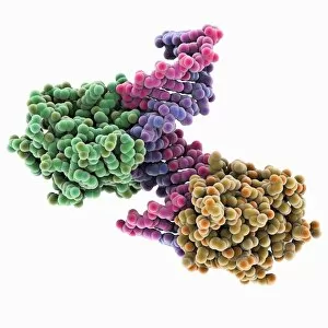
Oxoguanine glycosylase complex. Computer model showing a molecule of human aG DNA repair glycosylase (right) bound to an DNA molecule (left)

Background imageMolecules Collection: Green fluorescent protein molecule C013 / 8885

Green fluorescent protein molecule C013 / 8885
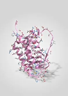
Green fluorescent protein molecule. Computer model showing the secondary structure of a molecule of green fluorescent protein (GFP). GFP is found in the Pacific jellyfish Aequorea victoria

Background imageMolecules Collection: Oxoguanine glycosylase complex C013 / 8884

Oxoguanine glycosylase complex C013 / 8884
Oxoguanine glycosylase complex. Computer model showing an 8-Oxoguanine glycosylase (OGG1) molecule (green) bound to a section of DNA (deoxyribonucleic acid, pink and blue)

Background imageMolecules Collection: Erythropoietin hormone complex

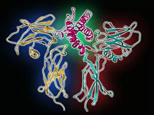
Erythropoietin hormone complex. Computer model showing the secondary structure of a molecule of the human hormone Erythropoietin (EPO), complexed with an erythropoetin receptor molecule

Background imageMolecules Collection: TATA box-binding protein complex C013 / 8881

TATA box-binding protein complex C013 / 8881
TATA box-binding protein complex. Molecular model showing a TATA box-binding protein (TBP) (purple) complexed with a strand of DNA (deoxyribonucleic acid, blue)

Background imageMolecules Collection: Bovine adrenodoxin molecules C013 / 8876

Bovine adrenodoxin molecules C013 / 8876
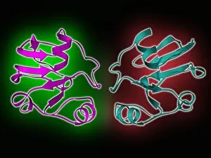
Bovine adrenodoxin molecules. Computer models showing the secondary structure of two molecules of adrenodoxin (adrenal ferredoxin) from a cow

Background imageMolecules Collection: Pit-1 transcription factor bound to DNA C013 / 8872

Pit-1 transcription factor bound to DNA C013 / 8872
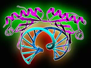
Pit-1 transcription factor bound to DNA. Molecular model showing pituitary-specific positive transcription factor 1 (Pit-1) (purple and yellow) bound to a strand of DNA (deoxyribonucleic acid)

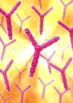
Background imageMolecules Collection: Antibodies, artwork C013 / 8800

Antibodies, artwork C013 / 8800
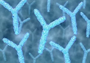
Antibodies. Computer artwork of antibody, or immunoglobulin, molecules. These y-shaped molecules have two arms that can bind to specific antigens, for instance viral or bacterial proteins

Background imageMolecules Collection: Tumour suppressor protein molecular model C013 / 7914

Tumour suppressor protein molecular model C013 / 7914
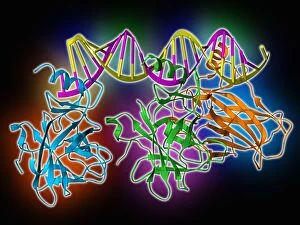
Tumour suppressor protein. Molecular model of the tumour suppressor protein p53 (blue, green and orange) bound to a molecule of DNA (deoxyribonucleic acid, yellow and pink)

Background imageMolecules Collection: DNA polymerase molecule C013 / 7909

DNA polymerase molecule C013 / 7909
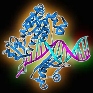
DNA polymerase. Molecular model of a molecule of DNA polymerase (blue) replicating a strand of DNA (deoxyribonucleic acid, pink and turquoise). The secondary structure of the DNA polymerase is shown

Background imageMolecules Collection: Antibodies, artwork C013 / 7792

Antibodies, artwork C013 / 7792
Antibodies. Computer artwork of antibody, or immunoglobulin, molecules. These y-shaped molecules have two arms that can bind to specific antigens, for instance viral or bacterial proteins

Background imageMolecules Collection: Antibodies, artwork C013 / 7791

Antibodies, artwork C013 / 7791
Antibodies. Computer artwork of antibody, or immunoglobulin, molecules. These y-shaped molecules have two arms that can bind to specific antigens, for instance viral or bacterial proteins

Background imageMolecules Collection: Antibodies, artwork C013 / 4683

Antibodies, artwork C013 / 4683
Antibodies. Computer artwork of antibody, or immunoglobulin, molecules. The y-shaped molecules have two arms that can bind to specific antigens, for instance viral or bacterial proteins

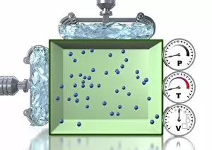
Background imageMolecules Collection: Boyles law of gases, artwork

Boyles law of gases, artwork
Boyles law of gases. Computer artwork explaining the gas laws as described by the combined gas law equation: PV=kT, which shows the relationship between the pressure (P), volume (V)

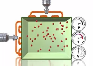
Background imageMolecules Collection: Combined gas law, artwork C013 / 4731

Combined gas law, artwork C013 / 4731
Combined gas law. Computer artwork explaining the gas laws as described by the combined gas law equation: PV=kT, which shows the relationship between the pressure (P), volume (V)

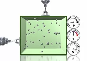
Background imageMolecules Collection: Pressure-temperature gas law, artwork C013 / 4730

Pressure-temperature gas law, artwork C013 / 4730
Pressure-temperature gas law. Computer artwork explaining the gas laws as described by the combined gas law equation: PV=kT, which shows the relationship between the pressure (P), volume (V)

Background imageMolecules Collection: Pressure-temperature gas law, artwork C013 / 4729

Pressure-temperature gas law, artwork C013 / 4729
Pressure-temperature gas law. Computer artwork explaining the gas laws as described by the combined gas law equation: PV=kT, which shows the relationship between the pressure (P), volume (V)

Background imageMolecules Collection: Viral recognition by antibodies, artwork C013 / 4722

Viral recognition by antibodies, artwork C013 / 4722
Viral recognition by antibodies. Computer artwork of rabies (family Rhabdoviridae) virus particles (virions, pink) being identified by monoclonal antibodies (Y-shaped, blue)

Background imageMolecules Collection: Atmospheric pressure explained, artwork C013 / 4712

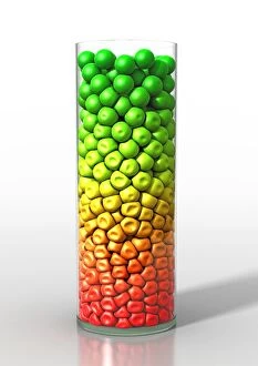
Atmospheric pressure explained, artwork C013 / 4712
Atmospheric pressure explained. Computer artwork of a number of small balls in a glass jar, representing the molecules present in a sample of liquid or gas

Background imageMolecules Collection: Aurora Borealis at night, Finland, february

Aurora Borealis at night, Finland, february

Background imageMolecules Collection: Aurora Borealis, over coniferous forest at night, Finland, february

Aurora Borealis, over coniferous forest at night, Finland, february

Background imageMolecules Collection: Red blood cells and molecules, artwork

Red blood cells and molecules, artwork
Red blood cells and drug molecules, computer artwork. Red blood cells (erythrocytes) are responsible for supplying tissues with oxygen and are the most abundant type of cell in the blood

Background imageMolecules Collection: White blood cells

White blood cells. Computer artwork of B lymphocytes, or B cells, each containing an antibody molecule. B cells mature in the bone marrow

Background imageMolecules Collection: Universal joint, computer model

Universal joint, computer model. This mechanical joint design, made entirely from carbon (turquoise) and hydrogen (grey) atoms, is an example of nanotechnology

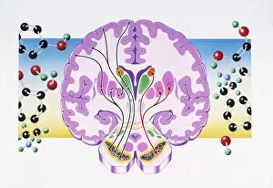
Background imageMolecules Collection: Brain drug

Brain drug

Background imageMolecules Collection: Human antibodies, TEM

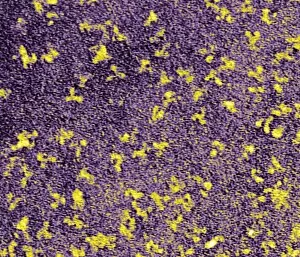
Human antibodies, TEM
Human antibodies (yellow), coloured transmission electron micrograph (TEM). The Y-shaped structures are molecules of the immunoglobulin G (IgG) antibody

Background imageMolecules Collection: Parkinsons disease

Parkinsons disease

Background imageMolecules Collection: Ozone molecules, artwork

Ozone molecules, artwork
Ozone molecules, computer artwork

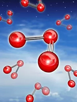
Background imageMolecules Collection: Oxygen molecule

Oxygen molecule, computer artwork. Molecular model of an oxygen molecule (O2). Two oxygen atoms (purple) are joined together. Oxygen is a colourless, odourless gas and is part of the chalcogen group

Background imageMolecules Collection: Gene therapy, artwork

Gene therapy, artwork
Gene therapy, conceptual computer artwork. Molecule of DNA (deoxyribonucleic acid, blue and orange), bound to a repair protein (purple), and a human figure

Background imageMolecules Collection: DNA with money

DNA with money. Computer artwork of a DNA double helix superimposed over a British 20 pound note. This image could represent the commercial implications of DNA research

Background imageMolecules Collection: Destruction of DNA helix

Destruction of DNA helix. The computer artwork may represent the breakdown of DNA during an explosion or the destruction of DNA in hot, molten lava

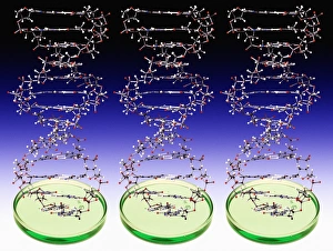
Background imageMolecules Collection: DNA molecules and Petri dishes

DNA molecules and Petri dishes
DNA molecules. Computer artwork of three molecules of DNA (deoxyribonucleic acid) emerging from three Petri dishes. DNA contains sections called genes that encode an organisms genetic information

Background imageMolecules Collection: Man and DNA

Man and DNA. Computer artwork of a DNA molecule wrapped around a male figure. A molecule of DNA (deoxyribonucleic acid) consists of two strands of sugar phosphates forming a double helix (spiral)


All Professionally Made to Order for Quick Shipping

"Molecules: The Building Blocks of Life and Beyond" From the intricate workings of an anaesthetic inhibiting an ion channel C015/6718 to the genius mind of James Clerk Maxwell, they have captivated scientists and artists alike. With their diverse structures and functions, they hold the key to understanding life at its core. Delving into the world of proteins, we witness their secondary structure through mesmerizing artwork that unveils their complexity. Meanwhile, the caffeine drug molecule keeps us awake while bacterial ribosomes tirelessly synthesize proteins within our cells. Vitamin B12's molecular model reminds us of nature's intricate design as zinc fingers elegantly bind to a DNA strand, orchestrating genetic processes. And who can forget capsaicin - the fiery molecule responsible for giving chili peppers their spicy kick? But molecules aren't limited to just earthly matters; they extend beyond our planet's boundaries. Oxytocin neurotransmitter molecules remind us of love's chemical connection while praziquantel parasite drugs combat infections in distant lands. Interferon molecules stand tall as defenders against viral invasions, showcasing our body's remarkable defense mechanisms. And amidst all this scientific wonder lies a breathtaking sight - Aurora Borealis dancing over a snow-covered coniferous forest in Northern Finland. Intricate and awe-inspiring, these glimpses into the molecular world remind us that there is so much more than meets the eye. From unlocking medical breakthroughs to unraveling nature's mysteries or simply marveling at captivating artistry – they can truly extraordinary entities shaping our understanding of life itself.