Fraud Blocker Skip to main content

Molecules Collection (page 2)

"Molecules

Background imageMolecules Collection: Carbon atom, digital illustration

Carbon atom, digital illustration

Background imageMolecules Collection: Aurora Borealis at night, Finland, January

Aurora Borealis at night, Finland, January

Background imageMolecules Collection: Aurora Borealis and stars over lake at night, Muonio, Lapland, Finland, September

Aurora Borealis and stars over lake at night, Muonio, Lapland, Finland, September

Background imageMolecules Collection: Aurora Borealis and stars over lake with beached canoes at night, Muonio, Lapland, Finland

Aurora Borealis and stars over lake with beached canoes at night, Muonio, Lapland, Finland, September

Background imageMolecules Collection: Aurora Borealis over fell at night, Saana Fell, Kilpisjarvi, Enontekio, Lapland, Finland, September

Aurora Borealis over fell at night, Saana Fell, Kilpisjarvi, Enontekio, Lapland, Finland, September

Background imageMolecules Collection: Aurora Borealis over lake with rowing boat at night, Lake Kilpisjarvi, Kilpisjarvi, Enontekio

Aurora Borealis over lake with rowing boat at night, Lake Kilpisjarvi, Kilpisjarvi, Enontekio, Lapland, Finland, September

Background imageMolecules Collection: Aurora Borealis and star trails over lake at night, Muonio, Lapland, Finland, September

Aurora Borealis and star trails over lake at night, Muonio, Lapland, Finland, September

Background imageMolecules Collection: Aurora Borealis, over coastline at night, Hornoya Island, Vardo, Finnmark, Norway, March

Aurora Borealis, over coastline at night, Hornoya Island, Vardo, Finnmark, Norway, March

Background imageMolecules Collection: Aurora Borealis, over taiga forest at night, Finland, January

Aurora Borealis, over taiga forest at night, Finland, January

Background imageMolecules Collection: Space-filling Models showing the Molecules in Decomposition of Sugar to Ethanol

Space-filling Models showing the Molecules in Decomposition of Sugar to Ethanol and Carbon Dioxide during Fermentation

Background imageMolecules Collection: Red-hot metal rod reaching white heat, close up

Red-hot metal rod reaching white heat, close up

Background imageMolecules Collection: Ball and Stick Model showing arrangement of Carbon Atoms in Diamond

Ball and Stick Model showing arrangement of Carbon Atoms in Diamond

Background imageMolecules Collection: Double Helix of Human DNA

Double Helix of Human DNA

Background imageMolecules Collection: Side view of a microscope with red and black wires

Side view of a microscope with red and black wires

Background imageMolecules Collection: Tumour suppressor protein and DNA C017 / 3647

Tumour suppressor protein and DNA C017 / 3647
Tumour suppressor protein and DNA. Computer artwork showing a molecule of the tumour suppressor protein p53 (blue and pink) bound to a molecule of DNA (deoxyribonucleic acid, yellow and orange)

Background imageMolecules Collection: Ricin A-chain, artwork C017 / 3653

Ricin A-chain, artwork C017 / 3653
Ricin A-chain. Computer artwork showing the enzymatically active A-chain from a molecule of the toxic protein ricin. Ricin comprises two entwined amino acid chains; A (seen here) and B (not shown)

Background imageMolecules Collection: Rotaxane, molecular crystal structure C017 / 7007

Rotaxane, molecular crystal structure C017 / 7007
Molecular crystal structure of a rotaxane. A rotaxane is a chemical compound composed of a linear molecular chain passing through a chainlike molecular ring

Background imageMolecules Collection: TATA box-binding protein complex C017 / 7082

TATA box-binding protein complex C017 / 7082
TATA box-binding protein complex. Molecular model showing a TATA box-binding protein (TBP, green) complexed with a strand of DNA (deoxyribonucleic acid, yellow) and transcription factor IIB

Background imageMolecules Collection: TATA box-binding protein complex C017 / 7088

TATA box-binding protein complex C017 / 7088
TATA box-binding protein complex. Molecular model showing a TATA box-binding protein (TBP, green) complexed with a strand of DNA (deoxyribonucleic acid, yellow) and transcription factor IIB

Background imageMolecules Collection: Ricin molecule, artwork C017 / 3652

Ricin molecule, artwork C017 / 3652
Ricin molecule. Computer artwork showing the structure of a molecule of the toxic protein ricin. Ricin comprises two entwined amino acid chains; A (yellow) and B (blue)

Background imageMolecules Collection: TATA box-binding protein complex C017 / 7084

TATA box-binding protein complex C017 / 7084
TATA box-binding protein complex. Molecular model showing a TATA box-binding protein (TBP, green) complexed with a strand of DNA (deoxyribonucleic acid, yellow) and transcription factor IIB

Background imageMolecules Collection: States of matter, artwork C017 / 3615

States of matter, artwork C017 / 3615
States of matter, computer artwork. From left to right the three states of matter are; solid, liquid and gas. Particles are in constant motion

Background imageMolecules Collection: Tumour suppressor protein and DNA C017 / 3644

Tumour suppressor protein and DNA C017 / 3644
Tumour suppressor protein and DNA. Computer artwork showing a molecule of the tumour suppressor protein p53 (blue and pink) bound to a molecule of DNA (deoxyribonucleic acid, yellow and orange)

Background imageMolecules Collection: Tunnelling current amplifier, artwork C017 / 3618

Tunnelling current amplifier, artwork C017 / 3618
Tunnelling current amplifier, computer artwork. Tunnelling current amplifiers are used in scanning tunnelling spectroscopy

Background imageMolecules Collection: Ricin molecule, artwork C017 / 3651

Ricin molecule, artwork C017 / 3651
Ricin molecule. Computer artwork showing the structure of a molecule of the toxic protein ricin. Ricin comprises two entwined amino acid chains; A (yellow) and B (blue)

Background imageMolecules Collection: Ricin molecule, artwork C017 / 3650

Ricin molecule, artwork C017 / 3650
Ricin molecule. Computer artwork showing the structure of a molecule of the toxic protein ricin. Ricin comprises two entwined amino acid chains; A (yellow) and B (blue)

Background imageMolecules Collection: Cytosine-guanine interaction, artwork C017 / 7215

Cytosine-guanine interaction, artwork C017 / 7215
Cytosine-guanine interaction. Computer artwork showing the structure of bound cytosine (left) and guanine molecules (right)

Background imageMolecules Collection: Tumour suppressor protein and DNA C017 / 3646

Tumour suppressor protein and DNA C017 / 3646
Tumour suppressor protein and DNA. Computer artwork showing a molecule of the tumour suppressor protein p53 (blue and pink) bound to a molecule of DNA (deoxyribonucleic acid, yellow and orange)

Background imageMolecules Collection: DNA components, artwork C017 / 7350

DNA components, artwork C017 / 7350
DNA components. Computer artwork showing the structure of the two molecules that make up the backbone of DNA (deoxyribonucleic acid), phosphate (left) and deoxyribose (right)

Background imageMolecules Collection: Cytosine-guanine interaction, artwork C017 / 7216

Cytosine-guanine interaction, artwork C017 / 7216
Cytosine-guanine interaction. Computer artwork showing the structure of bound cytosine (left) and guanine molecules (right)

Background imageMolecules Collection: Thymine-adenine interaction, artwork C017 / 7367

Thymine-adenine interaction, artwork C017 / 7367
Thymine-adenine interaction. Computer artwork showing the structure of bound thymine and adenine molecules. Atoms are shown as colour-coded spheres: carbon (green), hydrogen (white)

Background imageMolecules Collection: Circular DNA molecule, space artwork F006 / 7089

Circular DNA molecule, space artwork F006 / 7089
Circular DNA (deoxyribonucleic acid) molecule, computer artwork and space nebula artwork, depicting origin of life

Background imageMolecules Collection: Tablet computer, insulin molecule F006 / 6311

Tablet computer, insulin molecule F006 / 6311
Tablet computer showing a part of the molecule of human insulin. A single insulin molecule is made up of two chains of amino acids, the A and B chains, which are held together by di-sulphide bridges

Background imageMolecules Collection: Circular DNA molecule, space artwork F006 / 7077

Circular DNA molecule, space artwork F006 / 7077
Circular DNA (deoxyribonucleic acid) molecule, computer artwork and space nebula artwork, depicting origin of life

Background imageMolecules Collection: Circular DNA molecule, space artwork F006 / 7087

Circular DNA molecule, space artwork F006 / 7087
Circular DNA (deoxyribonucleic acid) molecule, computer artwork and space nebula artwork, depicting origin of life

Background imageMolecules Collection: Heat shock factor protein F007 / 9885

Heat shock factor protein F007 / 9885
Molecular model of a Heat Shock Protein (HSP).HSPs are a group of proteins whose levels increase when cells are exposed to raised temperatures or other stress

Background imageMolecules Collection: Heat shock factor 70 protein F007 / 9895

Heat shock factor 70 protein F007 / 9895
Molecular model of the Heat Shock Protein 70 (HSP).HSPs are a group of proteins whose levels increase when cells are exposed to raised temperatures or other stress

Background imageMolecules Collection: Haemagglutinin viral surface protein F007 / 9932

Haemagglutinin viral surface protein F007 / 9932
Haemagglutinin viral surface protein. Molecular model of haemagglutinin, a surface protein from the influenza virus, complexed with a neutralising antibody

Background imageMolecules Collection: Haemagglutinin viral surface protein F007 / 9931

Haemagglutinin viral surface protein F007 / 9931
Haemagglutinin viral surface protein. Molecular model of haemagglutinin, a surface protein from the influenza virus, complexed with a neutralising antibody

Background imageMolecules Collection: Heat shock factor 70 protein F007 / 9903

Heat shock factor 70 protein F007 / 9903
Molecular model of the Heat Shock Protein 70 (HSP).HSPs are a group of proteins whose levels increase when cells are exposed to raised temperatures or other stress

Background imageMolecules Collection: Human 80S ribosome F007 / 9902

Human 80S ribosome F007 / 9902
Ribosomal subunit. Computer model showing the structure of the RNA (ribonucleic acid) molecules in an 80S (large) ribosomal sub-unit. Ribosomes are composed of protein and RNA

Background imageMolecules Collection: Heat shock factor protein F007 / 9892

Heat shock factor protein F007 / 9892
Molecular model of a Heat Shock Protein (HSP).HSPs are a group of proteins whose levels increase when cells are exposed to raised temperatures or other stress

Background imageMolecules Collection: Human 80S ribosome F007 / 9898

Human 80S ribosome F007 / 9898
Ribosomal subunit. Computer model showing the structure of the RNA (ribonucleic acid) molecules in an 80S (large) ribosomal sub-unit. Ribosomes are composed of protein and RNA

Background imageMolecules Collection: Type I topoisomerase protein bound to DNA F007 / 9893

Type I topoisomerase protein bound to DNA F007 / 9893
Type I topoisomerase bound to DNA. Molecular model showing a type I topoisomerase molecule (blue) bound to a strand of DNA (deoxyribonucleic acid, yellow and red)

Background imageMolecules Collection: TATA box-binding protein complex C014 / 0867

TATA box-binding protein complex C014 / 0867
TATA box-binding protein complex. Molecular model showing a TATA box-binding protein (TBP, khaki) complexed with a strand of DNA (deoxyribonucleic acid)

Background imageMolecules Collection: E. coli Holliday junction complex C014 / 0878

E. coli Holliday junction complex C014 / 0878
E. coli Holliday junction complex. Molecular model of a RuvA protein (dark pink) in complex with a Holliday junction between homologous strands of DNA (deoxyribonucleic acid)

Background imageMolecules Collection: Iron containing protein, molecular model

Iron containing protein, molecular model
Iron containing protein. Molecular model showing the structure of a bacterial homolog of the animal iron containing protein ferritin

Background imageMolecules Collection: Streptavidin-biotin molecular complex

Streptavidin-biotin molecular complex. Molecular model of a single-strand binding complex of streptavidin (ribbons) and biotin (space-filled model, centre). Biotin is also known as vitamin B7


All Professionally Made to Order for Quick Shipping

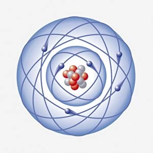
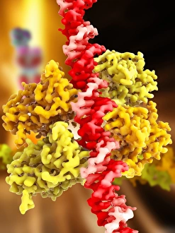
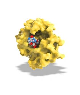
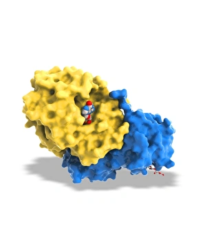
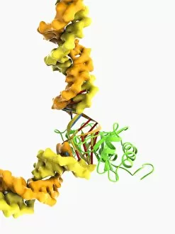
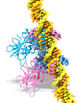
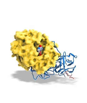
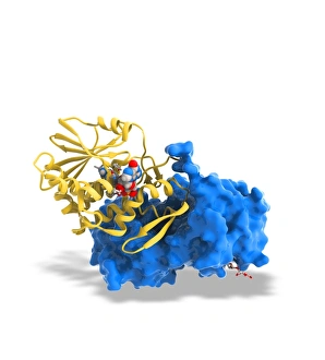
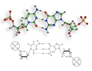
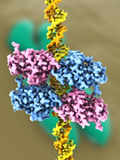
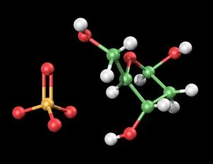
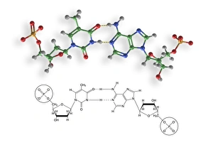
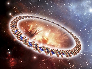
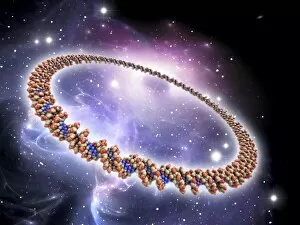
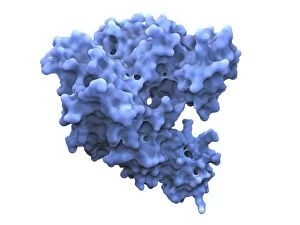
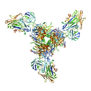
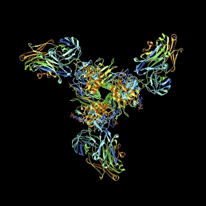
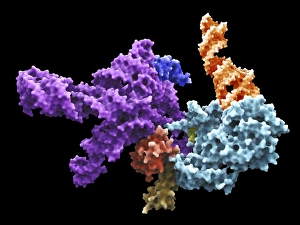
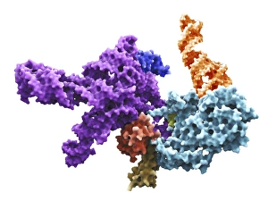
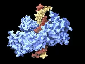
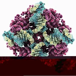
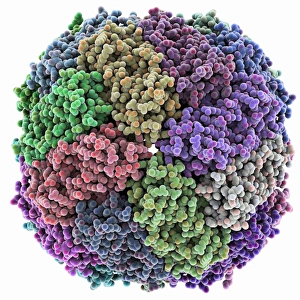
"Molecules: The Building Blocks of Life and Beyond" From the intricate workings of an anaesthetic inhibiting an ion channel C015/6718 to the genius mind of James Clerk Maxwell, they have captivated scientists and artists alike. With their diverse structures and functions, they hold the key to understanding life at its core. Delving into the world of proteins, we witness their secondary structure through mesmerizing artwork that unveils their complexity. Meanwhile, the caffeine drug molecule keeps us awake while bacterial ribosomes tirelessly synthesize proteins within our cells. Vitamin B12's molecular model reminds us of nature's intricate design as zinc fingers elegantly bind to a DNA strand, orchestrating genetic processes. And who can forget capsaicin - the fiery molecule responsible for giving chili peppers their spicy kick? But molecules aren't limited to just earthly matters; they extend beyond our planet's boundaries. Oxytocin neurotransmitter molecules remind us of love's chemical connection while praziquantel parasite drugs combat infections in distant lands. Interferon molecules stand tall as defenders against viral invasions, showcasing our body's remarkable defense mechanisms. And amidst all this scientific wonder lies a breathtaking sight - Aurora Borealis dancing over a snow-covered coniferous forest in Northern Finland. Intricate and awe-inspiring, these glimpses into the molecular world remind us that there is so much more than meets the eye. From unlocking medical breakthroughs to unraveling nature's mysteries or simply marveling at captivating artistry – they can truly extraordinary entities shaping our understanding of life itself.