Fraud Blocker Skip to main content

Molecules Collection (page 7)

"Molecules

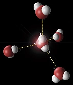
Background imageMolecules Collection: Hydrogen bonding in water, artwork C018 / 3560

Hydrogen bonding in water, artwork C018 / 3560
Hydrogen bonding in water. Artwork showing the hydrogen bonding (yellow dotted lines) between five water molecules. There is a weak, partial negative charge on the oxygen atoms (red)

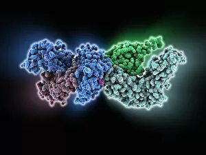
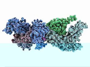
Background imageMolecules Collection: Human immune response molecule complex

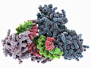
Human immune response molecule complex. Molecular model showing a human T-cell receptor and an HLA-A leukocyte (white blood cell) antigen bound to a TAX peptide from a virus

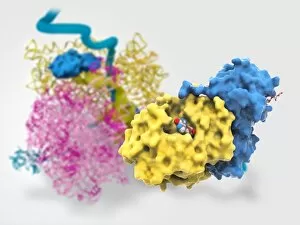
Background imageMolecules Collection: Ricin molecule, artwork C017 / 3648

Ricin molecule, artwork C017 / 3648
Ricin molecule Computer artwork showing the structure of a molecule of the toxic protein ricin (blue and yellow) with an active ribosome in the background

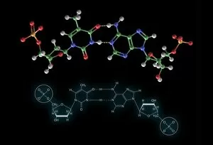
Background imageMolecules Collection: Thymine-adenine interaction, artwork C017 / 7368

Thymine-adenine interaction, artwork C017 / 7368
Thymine-adenine interaction. Computer artwork showing the structure of bound thymine and adenine molecules. Atoms are shown as colour-coded spheres: carbon (green), hydrogen (white)

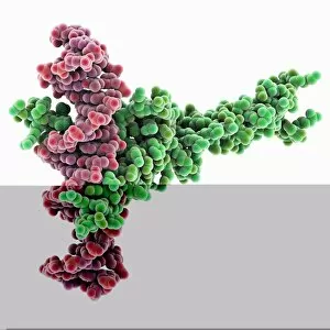
Background imageMolecules Collection: Pho4 transcription factor bound to DNA

Pho4 transcription factor bound to DNA. Molecular model showing phosphate system positive regulatory protein (Pho4) (pink and green) bound to a strand of DNA (deoxyribonucleic acid)

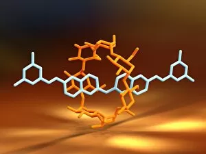
Background imageMolecules Collection: Rotaxane, molecular crystal structure C017 / 7010

Rotaxane, molecular crystal structure C017 / 7010
Molecular crystal structure of a rotaxane. A rotaxane is a chemical compound composed of a linear molecular chain passing through a chainlike molecular ring

Background imageMolecules Collection: TATA box-binding protein complex C017 / 7087

TATA box-binding protein complex C017 / 7087
TATA box-binding protein complex. Molecular model showing a TATA box-binding protein (TBP, green) complexed with a strand of DNA (deoxyribonucleic acid, yellow) and transcription factor IIB

Background imageMolecules Collection: Human immune response molecule complex C014 / 0871

Human immune response molecule complex C014 / 0871
Human immune response molecule complex. Molecular model showing a human T-cell receptor and an HLA-A leukocyte (white blood cell) antigen bound to a TAX peptide from a virus

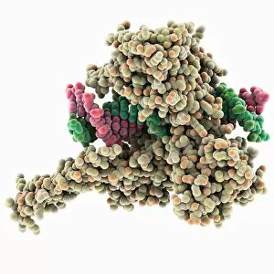
Background imageMolecules Collection: Type I topoisomerase bound to DNA C014 / 0862

Type I topoisomerase bound to DNA C014 / 0862
Type I topoisomerase bound to DNA. Molecular model showing a type I topoisomerase molecule (khaki) bound to a strand of DNA (deoxyribonucleic acid, pink and green)

Background imageMolecules Collection: Aspartyl-tRNA synthetase protein molecule C014 / 0874

Aspartyl-tRNA synthetase protein molecule C014 / 0874
Aspartyl-tRNA synthetase protein molecule. Molecular model showing the structure of the active site of aspartyl-tRNA synthetase (DARS) from yeast

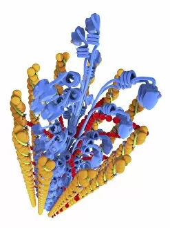
Background imageMolecules Collection: Actin Myosin Muscle Model, artwork C014 / 2658

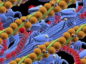
Actin Myosin Muscle Model, artwork C014 / 2658
Computer artwork of the molecular actin myosin muscle structure. The complex ultrastructure of cells, their shape and internal structure

Background imageMolecules Collection: Transcription factor bound to DNA C014 / 0868

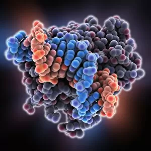
Transcription factor bound to DNA C014 / 0868
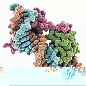
Transcription factor bound to DNA. Molecular model showing a MATa1/MATalpha2 homeodomain heterodimer (green and pink) in complex with a strand of DNA (deoxyribonucleic acid, orange and blue)

Background imageMolecules Collection: Amyloid precursor protein molecule

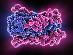
Amyloid precursor protein molecule
Amyloid precursor protein. Molecular model showing the structure of the protease inhibitor domain of an amyloid precursor protein (APP)

Background imageMolecules Collection: Amyloid precursor protein molecule C014 / 0863

Amyloid precursor protein molecule C014 / 0863
Amyloid precursor protein. Molecular model showing the structure of the protease inhibitor domain of an amyloid precursor protein (APP)

Background imageMolecules Collection: E. coli Holliday junction complex

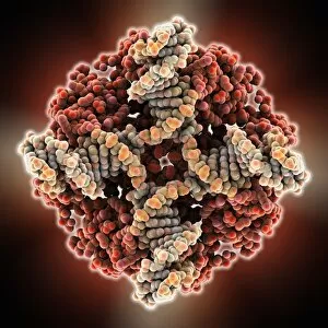
E. coli Holliday junction complex. Molecular model of a RuvA protein (red) in complex with a Holliday junction between homologous strands of DNA (deoxyribonucleic acid, brown and orange) from an E

Background imageMolecules Collection: TATA box-binding protein complex C014 / 0879

TATA box-binding protein complex C014 / 0879
TATA box-binding protein complex. Molecular model showing a TATA box-binding protein (TBP, lilac) complexed with a strand of DNA (deoxyribonucleic acid, green and red)

Background imageMolecules Collection: Methyladenine glycosylase bound to DNA C014 / 0877

Methyladenine glycosylase bound to DNA C014 / 0877
Methyladenine glycosylase bound to DNA. Computer model showing a molecule of human DNA-3-methyladenine glycosylase (purple) in complex with DNA (deoxyribonucleic acid, green and orange)

Background imageMolecules Collection: Type I topoisomerase bound to DNA C014 / 0883

Type I topoisomerase bound to DNA C014 / 0883
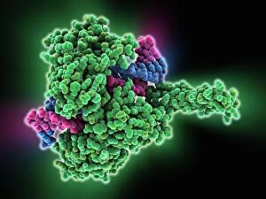
Type I topoisomerase bound to DNA. Molecular model showing a type I topoisomerase molecule (green) bound to a strand of DNA (deoxyribonucleic acid, pink and blue)

Background imageMolecules Collection: Actin Myosin Muscle Model, artwork C014 / 2659

Actin Myosin Muscle Model, artwork C014 / 2659
Computer artwork of the molecular actin myosin muscle structure. The complex ultrastructure of cells, their shape and internal structure

Background imageMolecules Collection: Actin myosin muscle model, artwork C014 / 2660

Actin myosin muscle model, artwork C014 / 2660
Computer artwork of the molecular actin myosin muscle structure. The complex ultrastructure of cells, their shape and internal structure

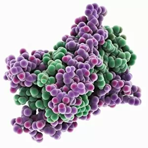
Background imageMolecules Collection: Methyladenine glycosylase bound to DNA

Methyladenine glycosylase bound to DNA. Computer model showing a molecule of human DNA-3-methyladenine glycosylase (purple) in complex with DNA (deoxyribonucleic acid, blue and orange)

Background imageMolecules Collection: HIV gp41 glycoprotein C014 / 0866

HIV gp41 glycoprotein C014 / 0866
HIV gp41 glycoprotein. Model showing the molecular structure of the gp41 protein from the HIV (human immunodeficiency virus) glycoprotein envelope

Background imageMolecules Collection: Zinc finger bound to DNA C014 / 0864

Zinc finger bound to DNA C014 / 0864
Zinc finger bound to DNA. Molecular model showing a zinc finger molecule bound to a strand of DNA (deoxyribonucleic acid)

Background imageMolecules Collection: Epstein-Barr virus protein bound to DNA C014 / 0875

Epstein-Barr virus protein bound to DNA C014 / 0875
Epstein-Barr virus protein bound to DNA. Computer model showing a molecule of Epstein-Barr nuclear antigen 1 (EBNA1) bound to a strand of DNA (deoxyribonucleic acid)

Background imageMolecules Collection: HIV gp41 glycoprotein

HIV gp41 glycoprotein. Model showing the molecular structure of the gp41 protein from the HIV (human immunodeficiency virus) glycoprotein envelope

Background imageMolecules Collection: ATP synthase molecule

ATP synthase molecule. Molecular model showing the structure of ATP synthase (ATPase) subunit C. ATPase is an important enzyme that provides energy for cells through the synthesis of adenosine

Background imageMolecules Collection: Transcription factor complexed with DNA C014 / 0870

Transcription factor complexed with DNA C014 / 0870
Transcription factor complexed with DNA. Computer model showing a max protein (green) bound to a strand of DNA (deoxyribonucleic acid, pink)

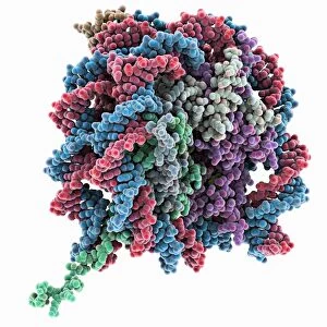
Background imageMolecules Collection: Nucleosome core particle bound to DNA C014 / 0872

Nucleosome core particle bound to DNA C014 / 0872
Nucleosome core particle bound to DNA. Molecular model showing a nucleosome core particle (green and purple) bound to a strand of DNA (deoxyribonucleic acid, blue and red)

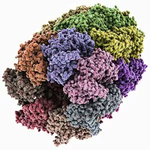
Background imageMolecules Collection: Chaperonin protein complex C014 / 0873

Chaperonin protein complex C014 / 0873
Chaperonin protein complex. Molecular model showing the structure of a GroEL/GroES/(ADP)7 chaperonin complex. Chaperonins are proteins that provide favourable conditions for the correct folding of

Background imageMolecules Collection: Chaperonin protein complex

Chaperonin protein complex. Molecular model showing the structure of a GroEL/GroES/(ADP)7 chaperonin complex. Chaperonins are proteins that provide favourable conditions for the correct folding of

Background imageMolecules Collection: DNA supercoils, artwork

DNA supercoils, artwork
DNA supercoils. Computer artwork showing DNA (deoxyribonucleic acid) in three stages of supercoiling. Supercoiling is important in a number of biological processes

Background imageMolecules Collection: Saliva chemicals, molecular model

Saliva chemicals, molecular model
Saliva chemicals. Molecular structure of a group of saliva molecules. These include the antibody immunoglobulin A (blue, double-y shape, see C014/5652)

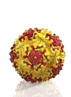
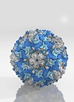
Background imageMolecules Collection: Enterovirus particle C014 / 4900

Enterovirus particle C014 / 4900
Enterovirus particle. Computer artwork of an enterovirus particle (virion), showing the structure of the capsid (outer shell)

Background imageMolecules Collection: Enterovirus capsid proteins structure C014 / 4897

Enterovirus capsid proteins structure C014 / 4897
Enterovirus capsid proteins structure. Computer artwork showing how the four component proteins (VP1 to VP4) of an enterovirus particle (virion) interlock to form the capsid (outer shell)

Background imageMolecules Collection: Enterovirus capsid proteins structure C014 / 4896

Enterovirus capsid proteins structure C014 / 4896
Enterovirus capsid proteins structure. Computer artwork showing how the four component proteins (VP1 to VP4) of an enterovirus particle (virion) interlock to form the capsid (outer shell)

Background imageMolecules Collection: Enterovirus particle C014 / 4898

Enterovirus particle C014 / 4898
Enterovirus particle. Computer artwork of an enterovirus particle (virion), showing the structure of the capsid (outer shell)

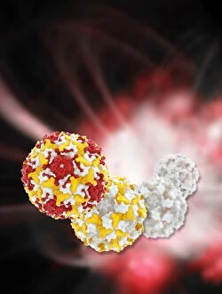
Background imageMolecules Collection: Enterovirus particles C014 / 4899

Enterovirus particles C014 / 4899
Enterovirus particles. Computer artwork of enterovirus particles (virion). Enteroviruses are a genus of non-enveloped positive-sense single-stranded RNA viruses associated with several human

Background imageMolecules Collection: UVR8 protein molecule C014 / 4912

UVR8 protein molecule C014 / 4912
UVR8 protein molecule. Computer model showing photoreception of UV-B (ultraviolet-B) light rays (top) by a UVR8 protein. The secondary structure (purple ribbons) of UVR8 is shown at bottom


All Professionally Made to Order for Quick Shipping

"Molecules: The Building Blocks of Life and Beyond" From the intricate workings of an anaesthetic inhibiting an ion channel C015/6718 to the genius mind of James Clerk Maxwell, they have captivated scientists and artists alike. With their diverse structures and functions, they hold the key to understanding life at its core. Delving into the world of proteins, we witness their secondary structure through mesmerizing artwork that unveils their complexity. Meanwhile, the caffeine drug molecule keeps us awake while bacterial ribosomes tirelessly synthesize proteins within our cells. Vitamin B12's molecular model reminds us of nature's intricate design as zinc fingers elegantly bind to a DNA strand, orchestrating genetic processes. And who can forget capsaicin - the fiery molecule responsible for giving chili peppers their spicy kick? But molecules aren't limited to just earthly matters; they extend beyond our planet's boundaries. Oxytocin neurotransmitter molecules remind us of love's chemical connection while praziquantel parasite drugs combat infections in distant lands. Interferon molecules stand tall as defenders against viral invasions, showcasing our body's remarkable defense mechanisms. And amidst all this scientific wonder lies a breathtaking sight - Aurora Borealis dancing over a snow-covered coniferous forest in Northern Finland. Intricate and awe-inspiring, these glimpses into the molecular world remind us that there is so much more than meets the eye. From unlocking medical breakthroughs to unraveling nature's mysteries or simply marveling at captivating artistry – they can truly extraordinary entities shaping our understanding of life itself.