
Mouth Part Collection
Exploring the Intricate World of Mouthparts: A Closer Look at Nature's Feeding Mechanisms Delicate and fascinating


All Professionally Made to Order for Quick Shipping
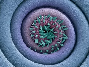
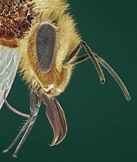
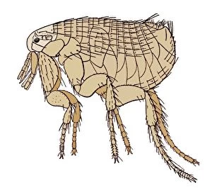
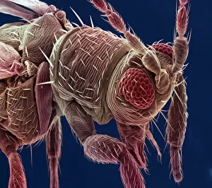
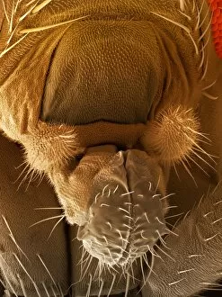
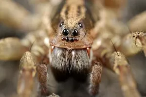
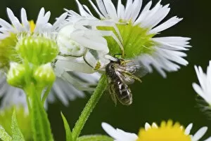
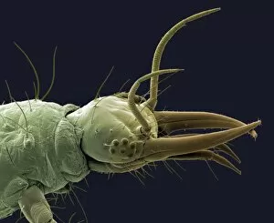
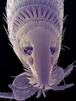
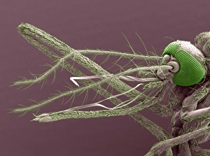
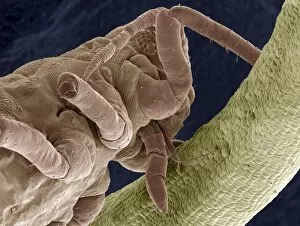
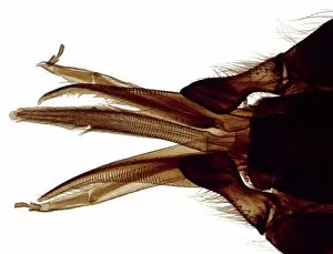
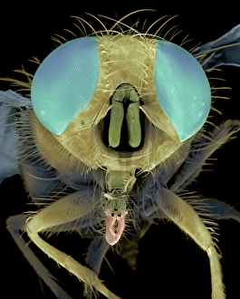
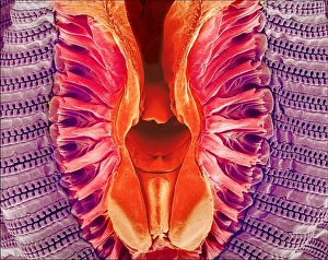
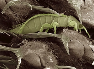
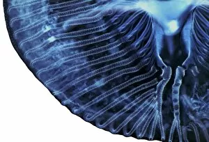
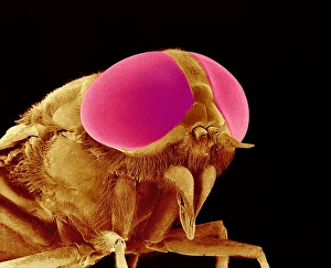
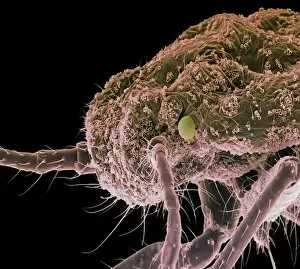
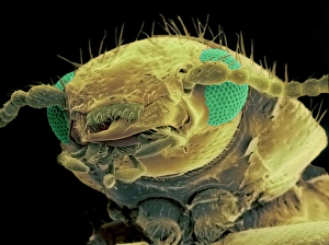
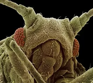
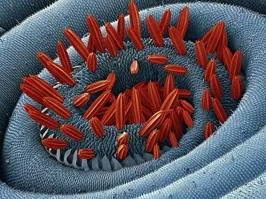
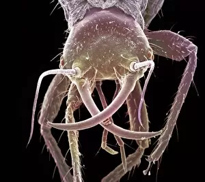
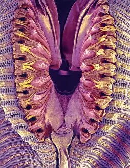
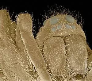
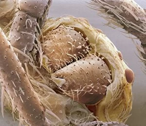
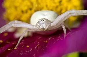
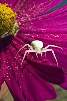
Exploring the Intricate World of Mouthparts: A Closer Look at Nature's Feeding Mechanisms Delicate and fascinating, the Moth proboscis reveals its intricate structure under the scanning electron microscope (SEM). Nature's design never ceases to amaze. The Head of a honey bee, captured through SEM, showcases its specialized mouthparts perfectly adapted for sipping nectar from flowers. Truly a marvel of evolution. An artwork depicting the Black Death rat flea reminds us of the historical significance these tiny creatures held as carriers of deadly diseases. Their menacing mouthparts played a crucial role in transmitting such devastating plagues. Magnified by SEM, the spiny spider's mouthpart appears like an intimidating weapon designed for capturing prey with precision and efficiency. Meet Thrip - a minuscule insect whose SEM image exposes its unique mouthpart structure responsible for piercing plant tissues and extracting sap. Tick mouthparts captured under SEM reveal their formidable nature – sharp hooks and barbs that enable them to latch onto hosts securely while feeding on blood. Honey bee head viewed through SEM offers an up-close look at their versatile mouthparts which aid in tasks ranging from collecting pollen to producing beeswax. Fruit fly proboscis magnified by SEM showcases its slender yet powerful tube-like structure used for probing fruits or other food sources with remarkable accuracy. 9/10. Wandering spiders C014 / 9804 & C014 / 9803 exhibit distinct variations in their impressive mandibles when observed under high-resolution microscopy - nature's diversity knows no bounds. Goldenrod crab spider captures a bee. Witnessing this dramatic moment emphasizes how crucial well-adapted mouthparts are for survival in the animal kingdom. SEM imagery unveils sheep tick’s complex set of cutting structures that allow it to attach firmly onto host animals while causing discomfort or transmitting diseases.