Fraud Blocker Skip to main content

Nasal Collection (page 2)

"Exploring the Nasal

Background imageNasal Collection: Facial anatomy, artwork

Facial anatomy, artwork
Facial anatomy. Computer artwork showing the superficial muscles around the nose and upper lip of a human face

Background imageNasal Collection: Facial hair clipper C014 / 1280

Facial hair clipper C014 / 1280
Facial hair clipper. Electric facial hair clipper for use on hair in places such as the ears, nose, and eyebrows

Background imageNasal Collection: Velvet skull, anatomical model

Velvet skull, anatomical model
Velvet skull. Velvet-covered anatomical model of a human skull

Background imageNasal Collection: Cooking smells, artwork C016 / 5343

Cooking smells, artwork C016 / 5343
Cooking smells. Artwork of two women inhaling cooking smells from a cooking pot

Background imageNasal Collection: Oestrus ovis, ship nasal botfly larva

Oestrus ovis, ship nasal botfly larva
A ship nasal botfly larva, a cause of myiasis extracted from a human eye in Kuwait. Photographed by Martin Hall

Background imageNasal Collection: Launch of the Space Shuttle Endeavour for STS-123 mission to the International Space Station

Launch of the Space Shuttle Endeavour for STS-123 mission to the International Space Station. John F. Kennedy Space Center in Cape Canaveral, Florida. March 11, 2008

Background imageNasal Collection: Anaesthetic Apparatus

Anaesthetic Apparatus
Frankis Evans Nasal Nitrous Oxide apparatus : (lower) Trewbys apparatus [This catalogue contains 2000 pages of medical equipment] Date: 1930

Background imageNasal Collection: Mole nose, SEM

Mole nose, SEM
Mole nose. Coloured scanning electron micrograph (SEM) of the snout (pink) of a mole, showing specialised bulbous papillae (projections) known as Eimers organs

Background imageNasal Collection: Nose, mouth and throat

Nose, mouth and throat. Cutaway artwork of the anatomy of the nose, mouth and throat. The large nasal cavity (upper centre) is lined with nerves (yellow) branching from the olfactory bulb (above)

Background imageNasal Collection: Nose mucosa, light micrograph

Nose mucosa, light micrograph
Nose mucosa. Light micrograph of a section through the nasal mucosa (the tissue that lines the airways of the nose), in the region lying over the nasal concha

Background imageNasal Collection: Illustration of nose & nasal mucosa

Illustration of nose & nasal mucosa

Background imageNasal Collection: Noses

Noses, artwork

Background imageNasal Collection: Skull, CT scan

Skull, CT scan
Human skull, coloured computed tomography (CT) scan. At top is the rounded cranium which houses the brain. The orbits (eye sockets) and nasal cavity (white) are seen

Background imageNasal Collection: Runny nose

Runny nose. Artwork of a section through the ear, nose and throat of a patient with excessive mucus production due to a cold or hay fever (allergic rhinitis)

Background imageNasal Collection: Nasal polyp, light micrograph

Nasal polyp, light micrograph
Nasal polyp. Light micrograph of a section through a nasal polyp, a benign growth that arises from a mucous membrane. Magnification: x150 when printed at 10 centimetres wide

Background imageNasal Collection: Nasal lining, TEM

Nasal lining, TEM
Nasal lining. Transmission electron micrograph (TEM) of a section through cilia (green) covering the epithelial lining of the nasopharyx, the part of the throat behind the nasal cavity

Background imageNasal Collection: Nasal lining, light micrograph

Nasal lining, light micrograph
Nasal lining. Light micrograph of a section through the olfactory epithelium. The olfactory epithelium is thicker than respiratory epithelium

Background imageNasal Collection: Hay fever, artwork

Hay fever, artwork
Hay fever. Artwork depicting hay fever showing pollen grains (yellow-blue) entering the nasal cavity (right) of a hay fever sufferer. Pollen grains are released when flowering plants reproduce

Background imageNasal Collection: Respiratory virus infection treatment

Respiratory virus infection treatment. Computer artwork showing virus particles (spherical) inside a persons nose and how they might be treated by inhaling a drug in the form of a nasal spray

Background imageNasal Collection: Human skull anatomy, artwork

Human skull anatomy, artwork
Human skull anatomy. Artwork showing the structure and anatomy of a human skull, including the sutures (jagged lines), the joints between the fused skull bones

Background imageNasal Collection: Nasal cavity and sinuses, artwork

Nasal cavity and sinuses, artwork
Nasal cavity and sinuses. Computer artwork of a coronal section through the face at the nose to show the nasal cavity and sinuses

Background imageNasal Collection: Head and neck anatomy, historical artwork

Head and neck anatomy, historical artwork. 19th Century hand coloured lithographic print showing the arteries (red) and muscle structure (brown) of the human neck and head

Background imageNasal Collection: Nose and brain anatomy, artwork

Nose and brain anatomy, artwork
Nose and brain anatomy. Artwork of a sagittal section through the human head, showing the anatomy of the brain and nasal cavity

Background imageNasal Collection: Nasal mucosa, artwork

Nasal mucosa, artwork
Nasal mucosa, cross-section. Artwork of a sequence (left to right) showing mucus production in response to infection and inflammation

Background imageNasal Collection: Head vascular anatomy, historical artwork

Head vascular anatomy, historical artwork. 19th Century hand coloured lithographic print showing the arteries (red) and muscle structure (brown) of the human neck and head

Background imageNasal Collection: Elderly woman blowing her nose

Elderly woman blowing her nose
MODEL RELEASED. Elderly woman blowing her nose with a tissue. Rhinitis (inflammation of the nose membranes) during a viral infection, leads to increased production of nasal mucus

Background imageNasal Collection: Skull bones, artwork

Skull bones, artwork
Skull bones. Computer artwork showing the major bones of the human skull. These are: frontal (purple, upper left), parietal (green/beige, upper right), occipital (blue, lower right), temporal (pink)

Background imageNasal Collection: Cartilage cells, TEM

Cartilage cells, TEM
Cartilage cells. Coloured transmission electron micrograph (TEM) of a section through chondrocytes from nasal hyaline cartilage

Background imageNasal Collection: Skull and eyeballs, artwork

Skull and eyeballs, artwork
Skull and eyeballs. Computer artwork of part of the skull, showing the eyeballs embedded in the eye sockets

Background imageNasal Collection: Nasal epithelium, SEM

Nasal epithelium, SEM
Nasal epithelium, coloured scanning electron micrograph (SEM). The nasal cavity contains both cilated (pink) and non-ciliated (secretory) cells

Background imageNasal Collection: A Nasal Reconaissance

A Nasal Reconaissance
Three British bobbies on the beat during WWI, attempt to lure a suspected German spy from his flat in Tottenham Court Road in London, by frying some delicious looking sausages beneath his window


All Professionally Made to Order for Quick Shipping

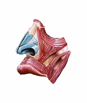
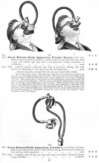
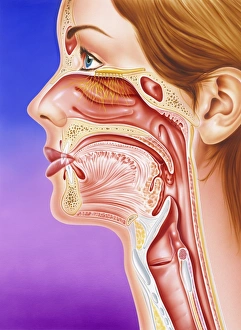
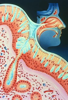
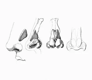
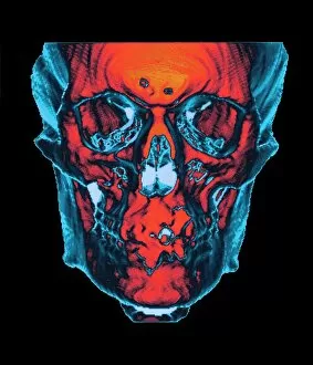
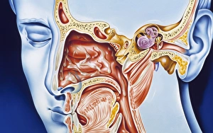
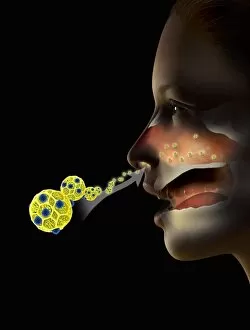
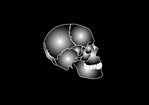
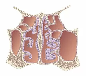
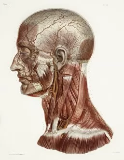
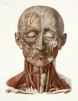
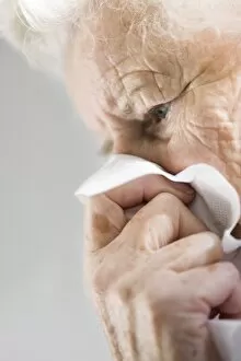
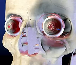
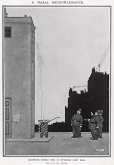
"Exploring the Nasal: Unveiling the Intricacies of Skull Anatomy by Leonardo da Vinci" Step into the world exploration as we delve into the fascinating realm of skull anatomy, guided by none other than Leonardo da Vinci. From horse skulls to human skulls, witness how this genius artist dissected and studied these intricate structures. Travel back in time to the 17th century and discover three distinct nose types that captivated artists and scientists alike. Marvel at a cutaway model of a face, revealing the hidden secrets beneath our skin. Uncover surprising connections between history and nasal protection as we showcase an iron hat worn by Charles I for nose defense. Journey further back in time with thirteenth-century cylindrical flat-topped helmets unearthed at Montgomery Castle, showcasing early forms protection. Witness how nasals have transcended mere functionality throughout centuries - from their role in ancient warfare to their presence in suffragette posters depicting women's struggles within prison walls. Immerse yourself in captivating visuals as a diagram overlays intricate nasal passages onto a human head. Admire Yadil Advertisement's artistic portrayal capturing the essence of beauty through highlighting delicate noses. Indulge your senses with Wishham Girl's photogravure from 1910, where every contour is beautifully captured, emphasizing the significance of nasals across cultures and eras. Join us on this enlightening journey through time as we unravel the mysteries surrounding one small yet vital feature -the nasal- leaving you astounded by its rich history and undeniable influence on art, science, fashion, and beyond.