Fraud Blocker Skip to main content

Nerve Cell Collection

The intricate world of nerve cells, also known as neurons, is a fascinating realm within our bodies

Background imageNerve Cell Collection: Cerebellum tissue, light micrograph

Cerebellum tissue, light micrograph
Cerebellum tissue. Confocal light micrograph of a section through the cerebellum of the brain. Purkinje cells, a type of neuron (nerve cell), are red

Background imageNerve Cell Collection: Purkinje nerve cells in the cerebellum

Purkinje nerve cells in the cerebellum
Purkinje cells in the cerebellum. Fluorescent light micrograph of Purkinje cells (green) in the cerebellum of the brain. Purkinje nerve cells have a flask-like body from which numerous highly

Background imageNerve Cell Collection: Nerve and glial cells, light micrograph

Nerve and glial cells, light micrograph
Nerve and glial cells, fluorescence light micrograph. These are neural stem cells that have differentiated into neurons (nerve cells, blue) and glial cells (support cells, red)

Background imageNerve Cell Collection: Hippocampus brain tissue

Hippocampus brain tissue
Hippocampus tissue. Light micrograph of a sagittal (side view) section through the hippocampus of the brain showing the nerve cells within it

Background imageNerve Cell Collection: Nerve cell, SEM

Nerve cell, SEM
Nerve cell. Coloured scanning electron micrograph (SEM) of a nerve cell (neuron). Neurons are responsible for passing information around the central nervous system (CNS)

Background imageNerve Cell Collection: Synapse nerve junction, TEM

Synapse nerve junction, TEM
Synapse. Coloured transmission electron micrograph (TEM) of a synapse, a junction between two nerve cells, in the brain. At a synapse an electrical signal is transmitted from one cell to the next in

Background imageNerve Cell Collection: Brain tissue blood supply

Brain tissue blood supply. Light micrograph of a section through cortex tissue from a brain, showing the blood vessels (branching) that supply it

Background imageNerve Cell Collection: Nerve cell

Nerve cell. Computer artwork of a nerve cell, also called a neuron. Neurons are responsible for passing information around the central nervous system (CNS) and from the CNS to the rest of the body

Background imageNerve Cell Collection: Cerebral cortex nerve cells

Cerebral cortex nerve cells. Confocal light micrograph of neurons (nerve cells, red) and glial cells (support cells, gold) from the cerebral cortex

Background imageNerve Cell Collection: Purkinje nerve cells in the cerebellum

Purkinje nerve cells in the cerebellum
Purkinje cells in the cerebellum. Fluorescent light micrograph of Purkinje cells (green) in the cerebellum of the brain. Purkinje nerve cells have a flask-like body from which numerous highly

Background imageNerve Cell Collection: Nerve cells

Nerve cells
Immunofluorescent Light Micrograph of neuron cells and astrocytes in mammalian spinal cord. Here, neuron cells stain red: the cell body appears pink

Background imageNerve Cell Collection: Cerebellum structure, light micrograph

Cerebellum structure, light micrograph
Cerebellum structure. Fluorescent light micrograph of a section through the cerebellum of the brain. The cerebellum comprises three main layers

Background imageNerve Cell Collection: Neural stem cell culture

Neural stem cell culture. Fluorescent light micrograph of a group of neural stem cells (neurosphere) in culture. Neural stem cells are able to differentiate into neurons (nerve cells)

Background imageNerve Cell Collection: Motor neurons, light micrograph

Motor neurons, light micrograph. Motor neurons are responsible for passing information around the central nervous system (CNS) and from the CNS to the rest of the body

Background imageNerve Cell Collection: Cerebellum tissue, light micrograph

Cerebellum tissue, light micrograph
Cerebellum tissue. Confocal light micrograph of a section through the cerebellum of the brain showing two types of glial cells (support cells); astrocytes (star-shaped)

Background imageNerve Cell Collection: Brain neuron

Brain neuron. Computer reconstruction of a medium spiny neuron from the basal ganglia of the brain. Neurons (nerve cells) are responsible for passing information around the central nervous system

Background imageNerve Cell Collection: Cerebellum tissue, light micrograph

Cerebellum tissue, light micrograph
Cerebellum tissue. Confocal light micrograph of a section through the cerebellum of the brain. Purkinje cells, a type of neuron (nerve cell), are red

Background imageNerve Cell Collection: Nerve cells, SEM

Nerve cells, SEM
Nerve cells. Coloured scanning electron micrograph (SEM) of nerve cells, known as neurones. Nerve cells occur in the brain, spinal cord, and in ganglia

Background imageNerve Cell Collection: Nerve cell, TEM

Nerve cell, TEM
Nerve cell. Coloured transmission electron micrograph (TEM) of a nerve cell body in cross- section. The cell has a large nucleus (yellow) and inner nucleolus (red)

Background imageNerve Cell Collection: Oligodendrocyte nerve cells

Oligodendrocyte nerve cells. Fluorescent light micrograph of human oligodendrocyte nerve cells. Cell nuclei, which contain the cells genetic information, have been dyed blue

Background imageNerve Cell Collection: Nerve cells, abstract artwork

Nerve cells, abstract artwork
Nerve cells. Abstract computer artwork of nerve cells, or neurons. Neurons are responsible for passing information around the central nervous system (CNS) and from the CNS to the rest of the body

Background imageNerve Cell Collection: Synapse nerve junctions, SEM

Synapse nerve junctions, SEM
Synapse nerve junctions. Coloured scanning electron micrograph (SEM) of nerve cells showing the synapses (junctions, bulges) between them

Background imageNerve Cell Collection: Nerve cell, artwork F007 / 7448

Nerve cell, artwork F007 / 7448
Nerve cell, computer artwork

Background imageNerve Cell Collection: Illustration of multipolar, unipolar, and bipolar neuron

Illustration of multipolar, unipolar, and bipolar neuron

Background imageNerve Cell Collection: Purkinje nerve cell, TEM C014 / 0583

Purkinje nerve cell, TEM C014 / 0583
Purkinje nerve cell. Transmission electron micrograph (TEM) of a purkinje nerve cell (green) from the cerebellum of the brain, showing the cell body (centre) and its primary dendrite (cell process)

Background imageNerve Cell Collection: Brain anatomy

Brain anatomy. Computer artwork of strands of nerve cells (neurons) in front of an exploded view of the brain seen from behind

Background imageNerve Cell Collection: Nerve synapse

Nerve synapse
Synapse. Illustration of a synaptic knob (at left), the junction between two nerve cells. When an electrical impulse reaches the synapse, vesicles (round blue shapes)

Background imageNerve Cell Collection: Astrocyte nerve cell

Astrocyte nerve cell. Fluorescent light micrograph of an astrocyte cell from a human brain. Intermediate filaments (IFs), part of the cells cytoskeleton, have been dyed green

Background imageNerve Cell Collection: Nerve synapse, TEM

Nerve synapse, TEM
Nerve synapse. Coloured transmission electron micrograph (TEM) of the neuron (nerve) terminal at a synapse in the diaphragm

Background imageNerve Cell Collection: Nerve cell, SEM

Nerve cell, SEM
Nerve cell. Coloured scanning electron micrograph (SEM) of a neuron (nerve cell). The cell body is the central structure with neurites (long and thin structures) radiating outwards from it

Background imageNerve Cell Collection: Nerve cells, computer artwork

Nerve cells, computer artwork
Nerve cells. Computer artwork of nerve cells, or neurons

Background imageNerve Cell Collection: Cross-section illustration of nasal cavity, nasal epithelium, and smell receptors (Olfaction)

Cross-section illustration of nasal cavity, nasal epithelium, and smell receptors (Olfaction)
Anatomy, Biomedical Illustration, Bone, Close-Up, Connection, Fragility, Human Hair, Human Head, Human Representation, Human Nose, Illustration and Painting, Illustrative Technique, Inside Of

Background imageNerve Cell Collection: Anterior cross section of spinal column revealing how vertebra fits round spinal cord

Anterior cross section of spinal column revealing how vertebra fits round spinal cord

Background imageNerve Cell Collection: Lateral cross section through the skull showing the brain with intermediate layers (meninges)

Lateral cross section through the skull showing the brain with intermediate layers (meninges) highlighted

Background imageNerve Cell Collection: Illustration of atom with nucleus of protons and neurons, based on the Bohr model

Illustration of atom with nucleus of protons and neurons, based on the Bohr model

Background imageNerve Cell Collection: Nerve synapse and serotonin molecule

Nerve synapse and serotonin molecule
Nerve synapse and serotonin neurotransmitter molecule. Computer artwork of a junction, or synapse, between two nerve cells (neurons, blue)

Background imageNerve Cell Collection: Neurology slide show C016 / 9008

Neurology slide show C016 / 9008
Conceptual computer artwork depicting neurology. From left to right: MRI brain scans, 3D dsi white matter brain scan, brain, Alzheimers brain versus normal brain, MRI brain scan

Background imageNerve Cell Collection: Cross-section diagram of human skin

Cross-section diagram of human skin

Background imageNerve Cell Collection: Illustration of Oligodendrocyte glia cell or neuroglia

Illustration of Oligodendrocyte glia cell or neuroglia

Background imageNerve Cell Collection: Digital illustration showing sensory neurons sending message to nerves in human arm after scalding b

Digital illustration showing sensory neurons sending message to nerves in human arm after scalding b

Background imageNerve Cell Collection: Digital illustration of a neuron

Digital illustration of a neuron

Background imageNerve Cell Collection: Digital illustration of of human nervous system

Digital illustration of of human nervous system

Background imageNerve Cell Collection: Cross section illustration of Synapse

Cross section illustration of Synapse

Background imageNerve Cell Collection: Cross section illustration of human skin showing touch receptor nerves

Cross section illustration of human skin showing touch receptor nerves

Background imageNerve Cell Collection: Illustration of different types of cells, nerve cells, red blood cell, muscle cell

Illustration of different types of cells, nerve cells, red blood cell, muscle cell, and guard cells of a leaf

Background imageNerve Cell Collection: Biomedical illustration of neural network at 6 years of age

Biomedical illustration of neural network at 6 years of age

Background imageNerve Cell Collection: Cross section biomedical illustration of nerves development with myelin winding around axons to

Cross section biomedical illustration of nerves development with myelin winding around axons to form protective and insulating sheath

Background imageNerve Cell Collection: Cross section biomedical illustration of nerve development with axon wrapped in sections of myelin

Cross section biomedical illustration of nerve development with axon wrapped in sections of myelin separated by Node of Ranvier


All Professionally Made to Order for Quick Shipping

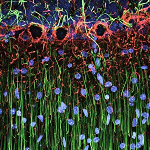
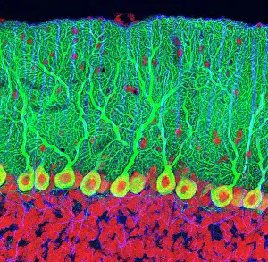
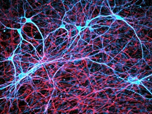
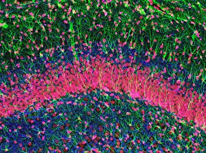
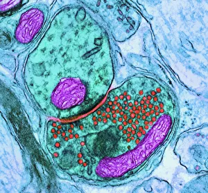
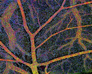
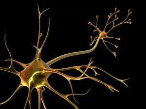
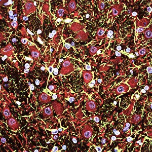
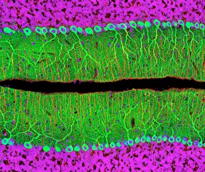
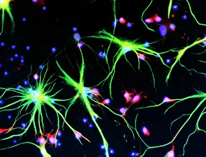
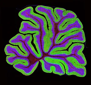
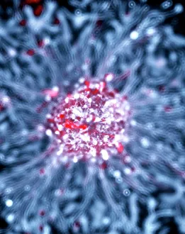
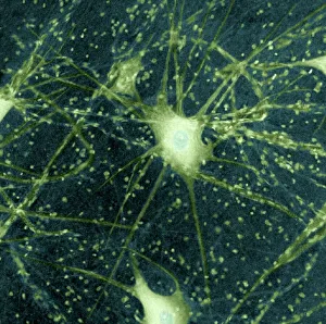
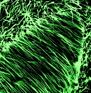
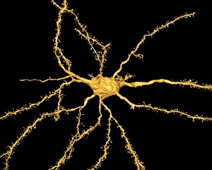
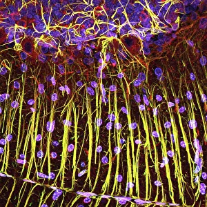
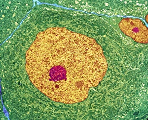
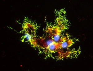
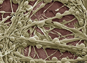
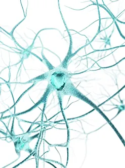
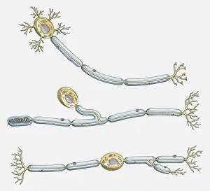
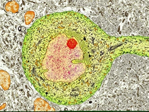
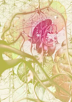
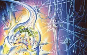
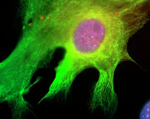
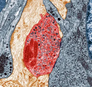
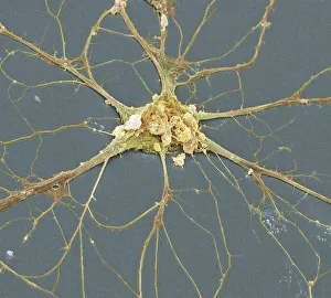
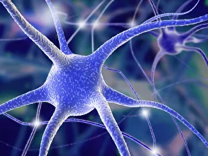
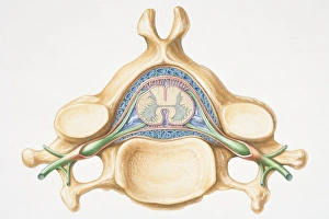
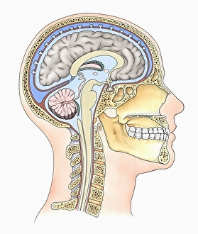
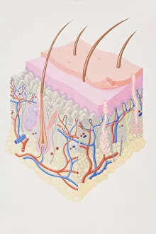
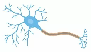
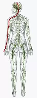
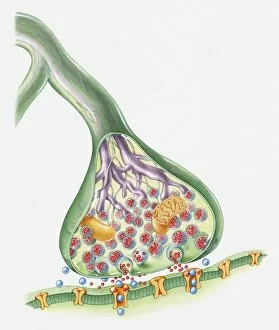
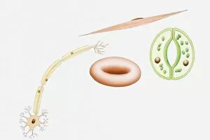
The intricate world of nerve cells, also known as neurons, is a fascinating realm within our bodies. These specialized cells play a crucial role in transmitting information throughout the nervous system. In the cerebellum tissue, under the lens of a light micrograph, we can observe the complex network formed by nerve and glial cells. This interplay between different cell types ensures proper functioning and communication within this region of the brain. Zooming in further with a transmission electron microscope (TEM), we witness the mesmerizing synapse nerve junctions - where two nerve cells meet to exchange vital signals. The intricacy of these connections highlights their importance in relaying messages across our neural pathways. Switching gears to scanning electron microscopy (SEM), we get an up-close look at individual nerve cells themselves. Their elongated structures and branching extensions showcase their ability to transmit electrical impulses efficiently. Moving on to hippocampus brain tissue, another essential area for memory formation and learning, we encounter Purkinje nerve cells nestled within the cerebellum. These large neurons have distinctive dendritic trees that receive inputs from various sources, contributing to motor coordination. As we explore further into brain tissue's complexity, it becomes evident that blood supply plays a crucial role in nourishing these delicate neural networks. A healthy flow ensures optimal functioning of all interconnected regions. Venturing beyond natural tissues into neural stem cell culture reveals exciting possibilities for regenerative medicine and understanding neurodevelopmental processes better. These cultured stem cells hold immense potential for repairing damaged nerves or studying neuronal growth patterns. Finally, let us not forget about cerebral cortex nerve cells - responsible for higher cognitive functions such as perception and decision-making. Their intricate arrangements enable us to process information effectively while navigating through daily life challenges. Whether observing cerebellum tissue or exploring neural stem cell cultures or marveling at synaptic connections under various microscopes – each glimpse into the world of nerve cells unveils new layers of complexity and highlights their indispensable role in our intricate neural symphony.