
Neuroglia Collection
Neuroglia, also known as glial cells, are the unsung heroes of our nervous system. These remarkable cells play a crucial role in supporting and protecting our neurons


All Professionally Made to Order for Quick Shipping
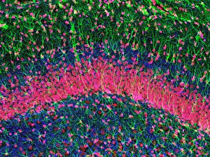
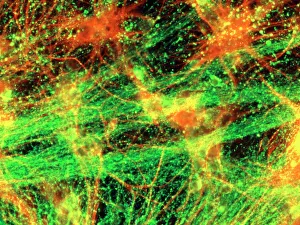
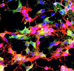
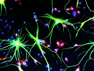
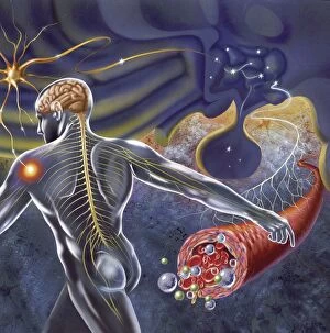
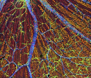
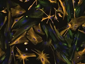
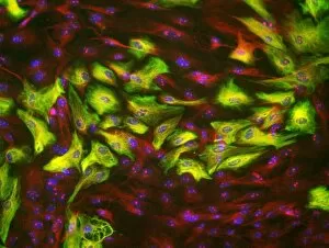
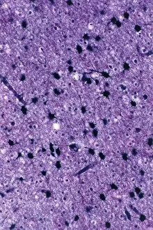
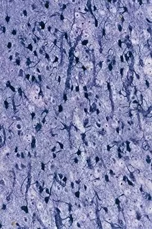
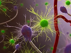
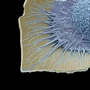
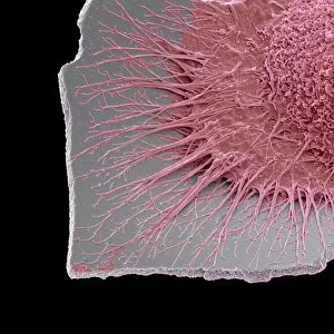
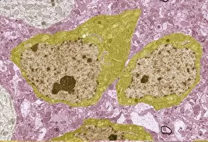
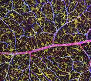
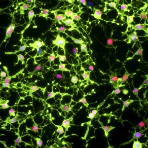
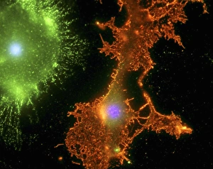
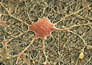
Neuroglia, also known as glial cells, are the unsung heroes of our nervous system. These remarkable cells play a crucial role in supporting and protecting our neurons. In an immunofluorescent light micrograph of neurons and astrocytes, we can see the intricate network they form within the hippocampus brain tissue. The vibrant colors highlight their presence amidst the nerve cells, emphasizing their importance. Moving to a glial stem cell culture captured under a light microscope, we witness these versatile cells in action. Their ability to differentiate into various types of glial cells is truly awe-inspiring. A scanning electron microscope image reveals another aspect of neuroglia's work - safeguarding our brain lining. In this SEM image, we observe how these specialized cells create a protective barrier that shields our precious neural tissue from harm. But neuroglia's responsibilities extend beyond just the brain; they even lend their support to other parts of our body. As shown in an SEM image of fallopian tube cells, these glial cells ensure proper functioning and communication throughout different systems. Delving deeper into their structure, a cross-section diagram illustrates how neuroglia interact with nerve fibers and blood vessels while enveloping them with myelin sheaths. This complex arrangement facilitates efficient transmission of nerve impulses throughout our body. An illustration showcases one specific type - oligodendrocyte glia cell. These unique structures wrap around axons forming myelin sheaths that enhance signal conduction speed along nerves. Zooming out for a broader view, a schematic depicts how the hypothalamus receives vital nerve impulses from various regions within our body. Neuroglia assist in processing this information accurately so that appropriate responses can be generated by other parts of the brain or organs. Another fascinating aspect is revealed through an image where we see a nerve connecting with muscle fibers via its myelin sheath at its lower right end.