
Nodes Collection
"Exploring the Intricate Network of Nodes: From Cardiovascular System to Historical Artwork" Delve into the fascinating world of nodes


All Professionally Made to Order for Quick Shipping
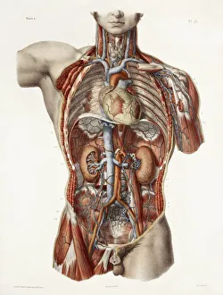
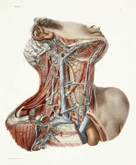
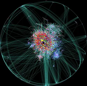
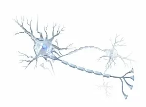
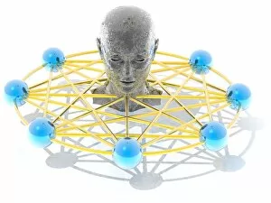
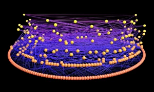
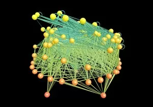
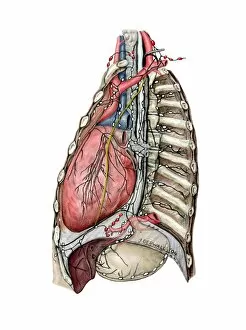
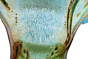
"Exploring the Intricate Network of Nodes: From Cardiovascular System to Historical Artwork" Delve into the fascinating world of nodes, where connections and pathways intertwine across various realms. In the cardiovascular system, nodes play a crucial role in regulating heartbeats and maintaining rhythm, ensuring our bodies function harmoniously. Just like historical artwork capturing moments frozen in time, these nodes serve as windows into our past, revealing tales of human anatomy. Shifting our focus to neck anatomy, we uncover an intricate web that facilitate communication between different body parts. As 19th-century artwork immortalizes this complexity on canvas, we gain a deeper appreciation for the marvels hidden beneath our skin. Venturing further into the digital age, imagine an internet blog map resembling a vast network of interconnected nodes. Each node represents a unique piece of information waiting to be discovered by curious minds navigating through cyberspace. However, not all nodal journeys are pleasant; some lead us down darker paths such as elephantiasis. X-ray images reveal how swollen lymph they are cause immense discomfort and hinder mobility – reminding us of the importance of early detection and treatment. Yet amidst these challenges lie moments of beauty – morning dew glistening on leaves like tiny droplets forming delicate networks connecting nature's elements. These transient yet enchanting formations remind us that even in simplicity lies intricacy worth cherishing. Returning to medical terrain once more with arthritic hands depicted through x-rays – bony protrusions resembling gnarled branches showcase how joints become entangled over time. Yet within this visual representation lies hope for understanding and managing this condition effectively. Zooming out from individual ailments towards nerve cells portrayed artistically reveals their mesmerizing complexity - each cell serving as a vital node transmitting signals throughout our bodies' neural networks. Finally, let's appreciate the grandeur encompassed within human lymphatic system artwork - showcasing its extensive reach from head to chest and deep into the back of our legs.