Fraud Blocker Skip to main content

Nodule Collection

In the quaint town of Buxton, England, lies a hidden treasure known as hematite nodules

Background imageNodule Collection: Hematite, Buxton, England

Hematite, Buxton, England
Massive, compact, water worn hematite, dark red in colour. The specimen label states Iron Ore, the best pieces are used for burnishing when polished - from Buxton or thereabouts

Background imageNodule Collection: Agate nodules

Agate nodules
6337602 Agate nodules by Corsi (19th century); (add.info.: Agate nodules. Ovoid nodule of agate 1, aphanite with agate geode 2, and nodule of agate 3)

Background imageNodule Collection: Seliguea feei fern

Seliguea feei fern. Selliguea del Fee, Selliguea fei. Handcoloured copperplate stipple engraving from Antoine Laurent de Jussieus Dizionario delle Scienze Naturali, Dictionary of Natural Science

Background imageNodule Collection: Diseases of the Skin: Rodent Ulcer and Epithelioma (colour litho)

Diseases of the Skin: Rodent Ulcer and Epithelioma (colour litho)
6015814 Diseases of the Skin: Rodent Ulcer and Epithelioma (colour litho) by English School, (19th century); Private Collection; (add.info.: Rodent Ulcer and Epithelioma)

Background imageNodule Collection: Humpback whale fluke surfacing in the water of the Southern Ocean, Antarctic Peninsula, Antarctica

Humpback whale fluke surfacing in the water of the Southern Ocean, Antarctic Peninsula, Antarctica
The tail of a humpback whale, Megaptera novaeangliae, off the Antarctic Peninsula

Background imageNodule Collection: Two humpback whales surfacing the water of the Southern Ocean off of Antarctica

Two humpback whales surfacing the water of the Southern Ocean off of Antarctica
Close-up portrait of two humpback whales (Megaptera novaeangliae) surfacing and splashing in the water of the Southern Ocean off of Antarctica; Southern Ocean, Antarctica

Background imageNodule Collection: Root nodule

Root nodule. Coloured scanning electron micrograph (SEM) of a root nodule on a pea plant (Pisum sativum) caused by the nitrogen-fixing soil bacteria Rhizobium leguminosarum

Background imageNodule Collection: Septarian concretion or Septarian Nodule in claystone, close-up

Septarian concretion or Septarian Nodule in claystone, close-up

Background imageNodule Collection: Picture No. 11050190

Picture No. 11050190
Cross-Section Utah Septarian Nodule Utah, USA Date:

Background imageNodule Collection: Minerals, Iron Pyrite or Fools Gold crystals, cubic crystals on host rock

Minerals, Iron Pyrite or Fools Gold crystals, cubic crystals on host rock

Background imageNodule Collection: Amber nodule

Amber nodule

Background imageNodule Collection: Pteridosperms - Paripteris: Compression fossil in ironstone nodule

Pteridosperms - Paripteris: Compression fossil in ironstone nodule

Background imageNodule Collection: Cirrhosis of the liver, light micrograph

Cirrhosis of the liver, light micrograph
Cirrhosis of the liver. Light micrograph of a section through liver tissue affected by cirrhosis, showing a cluster of hepatocyte cells (orange)

Background imageNodule Collection: Trilobite Assemblage

Trilobite Assemblage
Nodule containing a concentration of Ellipsocephalus hoffi (Schlotheim 1823) Middle Cambrian, Jince Formation, Hydrocephalus lyelli zone, Mount Konicek, Jince, Czech Republic. Pete Lawrance Collection

Background imageNodule Collection: Rock concretions and minerals C016 / 4850

Rock concretions and minerals C016 / 4850
Rock concretions and minerals. Clockwise from upper left: septarian nodule, fractured flint nodule, fractured pyrite nodule and a selenite desert rose

Background imageNodule Collection: Sectioned nitrogen-fixing nodule C013 / 7351

Sectioned nitrogen-fixing nodule C013 / 7351
Nitrogen-fixing nodules on roots of a tree lupin, Lupinus arboreus, caused by Rhizobium bacteria. The association between Rhizobium species and leguminous plants is an example of symbiosis

Background imageNodule Collection: Section through an iron meteorite C013 / 6567

Section through an iron meteorite C013 / 6567
Section through an iron meteorite. Iron meteorites, when sliced open and etched with acid, typically show a distinctive criss-cross pattern known as the Widmanstatten pattern

Background imageNodule Collection: Lymph node, artwork C013 / 4632

Lymph node, artwork C013 / 4632
Lymph node. Computer artwork of a section through a human lymph node. Lymph nodes are kidney-shaped structures located at intervals along lymph vessels (tubes)

Background imageNodule Collection: Lymph node, artwork C013 / 4631

Lymph node, artwork C013 / 4631
Lymph node. Computer artwork of a section through a human lymph node. Lymph nodes are kidney-shaped structures located at intervals along lymph vessels (tubes)

Background imageNodule Collection: Nitrogen-fixing root nodule, micrograph

Nitrogen-fixing root nodule, micrograph
Nitrogen-fixing root nodule. Light micrograph of a section through a root nodule of a broad bean plant (Faba vulgaris) showing the symbiotic Rhizobium leguminosarum bacteria (purple) within it

Background imageNodule Collection: Nitrogen-fixing bacteria

Nitrogen-fixing bacteria. Coloured scanning electron micrograph (SEM) of Rhizobium leguminosarum nitrogen fixing bacteria (brown)

Background imageNodule Collection: Precious opal in ironside nodule

Precious opal in ironside nodule
Opals are not truely crystalline and are therefore mineraloids. They comprise of (hydrated silica glass). Specimen found in Queensland, Australia. On display at the Natural History Museum, London

Background imageNodule Collection: Marcasite

Marcasite comprises of (iron sulphide). It is similar in appearance to pyrite, but has a different structural composition

Background imageNodule Collection: Wardite

Wardite is comprised of hydrated sodium aluminum phosphate hydroxide. This bright green specimen has been deposited in variscite nodules

Background imageNodule Collection: Ankylosaur skin nodule

Ankylosaur skin nodule
This nodule would have been attached to the dinosaurs back by its flat base with the broad ridge providing protection. The Ankylosaurs were a family of dinosaurs characterised by thick bony plated

Background imageNodule Collection: Shale

Shale
A specimen of shale containing small nodules of clay ironstone from the Lower Coal Measures, Brynamman, Carmarthenshire

Background imageNodule Collection: Concretions

Concretions

Background imageNodule Collection: Fossilised millipede (Class Diplopoda)

Fossilised millipede (Class Diplopoda)
This fossil is preserved in a siltstone nodule of Carboniferous age from the Yorkshire Coalfield. Length 63mm (unstraightened), length of nodule 76mm

Background imageNodule Collection: Septarian nodule

Septarian nodule

Background imageNodule Collection: Polacanthus skin nodule

Polacanthus skin nodule
These nodules were mixed in with the overlapping plates on Polacanthus skin similar to those in modern reptiles. Polacanthus lived 132 to 100 million years ago during the lower Cretaceous period

Background imageNodule Collection: Paraisobuthus prantli, scorpion

Paraisobuthus prantli, scorpion
A Scorpion fossil seen here in a nodule of rock

Background imageNodule Collection: Opal

Opal
Boulder or nodular opal in a sandstone matrix from Queensland, Australia. Opals are not truely crystalline and are therefore mineraloids. They comprise of (hydrated silica glass)

Background imageNodule Collection: Potentilla nivea L. snow cinquefoil

Potentilla nivea L. snow cinquefoil
Sketch 2, Newfoundland Volumes. From a collection of original drawings and sketches by Georg Dionysius Ehret (1708-1770). Held in the Botany Library at the Natural History Museum, London

Background imageNodule Collection: Flint nodule

Flint nodule
This flint nodule originates from the Cretaceous rocks of the North Downs, England

Background imageNodule Collection: Tonsil, light micrograph

Tonsil, light micrograph
Tonsil. Light micrograph of a section through the surface of a tonsil, showing a lymphoid nodule (purple). Tonsils are lymph nodes, which filter pathogens from tissue fluid

Background imageNodule Collection: Sarcoidosis of the lungs, X-ray

Sarcoidosis of the lungs, X-ray
Sarcoidosis. Frontal coloured chest X-ray of the lungs of a patient with pulmonary sarcoidosis. Affected lung tissue shows up as red patches

Background imageNodule Collection: Lymph node, light micrograph

Lymph node, light micrograph
Lymph node. Coloured light micrograph of a section through a lymph node. A lymph node filters pathogens from lymph fluid, a watery liquid that surrounds the tissues of the body

Background imageNodule Collection: Typhoid nodules, light micrograph

Typhoid nodules, light micrograph
Typhoid nodules, coloured light micrograph. Section through a lymph node of a patient with typhoid, showing macrophages (large cells, pink) forming typhoid nodules

Background imageNodule Collection: Liver tuberculosis, light micrograph

Liver tuberculosis, light micrograph
Liver tuberculosis. Coloured light micrograph of a section through the liver of a patient with miliary tuberculosis (TB). A tubercle, a nodular lesion of infected dead tissue, is seen at left

Background imageNodule Collection: Miliary tuberculosis, light micrograph

Miliary tuberculosis, light micrograph
Miliary tuberculosis. Light micrograph of a section through a single miliary tubercle (left) in a patients lung. Tubercles are nodular lesions of infected dead tissue that arise from tuberculosis (TB)

Background imageNodule Collection: Lymph node, TEM

Lymph node, TEM
Lymph node. Coloured transmission electron micrograph (TEM) of a section through the peripheral corona of a lymph nodule, showing numerous small lymphocyte white blood cells (dark)

Background imageNodule Collection: Tonsil tissue, light micrograph

Tonsil tissue, light micrograph

Background imageNodule Collection: Slice of agate, a form of quartz

Slice of agate, a form of quartz
Agate. Slice through a nodule of agate, a type of chalcedony. This is a cryptocrystalline variety of the mineral quartz (silicon oxide)

Background imageNodule Collection: Nitrogen-fixing bacteria, SEM

Nitrogen-fixing bacteria, SEM
Nitrogen-fixing bacteria. Coloured scanning electron micrograph (SEM) of nitrogen-fixing soil bacteria (Rhizobium sp.) living on a root nodule of a runner bean plant (Phaseolus coccineus)

Background imageNodule Collection: Legume root nodules, light micrograph

Legume root nodules, light micrograph
Legume root nodules. Light micrograph of sections through two root nodules from a broad bean plant (Vicia faber). These root nodules are full of nitrogen-fixing bacteria

Background imageNodule Collection: Broad bean root nodules, light micrograph

Broad bean root nodules, light micrograph
Broad bean root nodules. Light micrograph of the sections of the root nodules of the broad bean (Vicia faber) which are full of nitrogen-fixing bacteria


All Professionally Made to Order for Quick Shipping

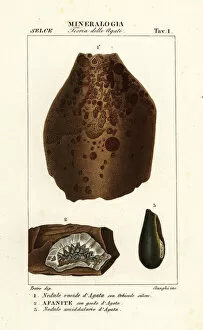
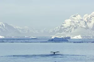
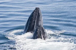
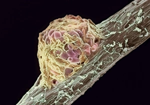
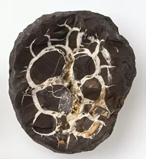
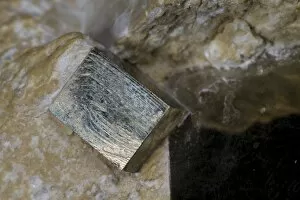
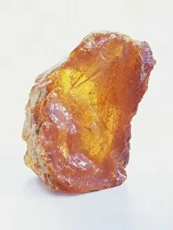
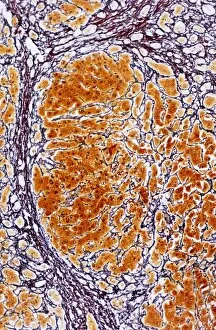
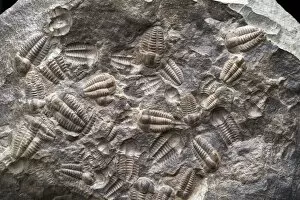
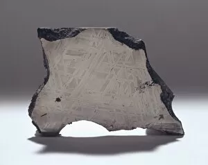
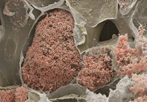
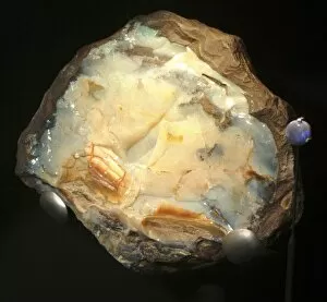
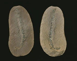
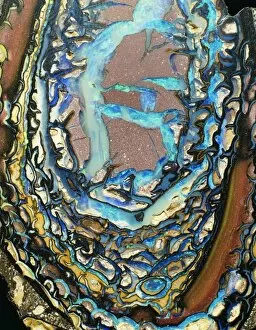
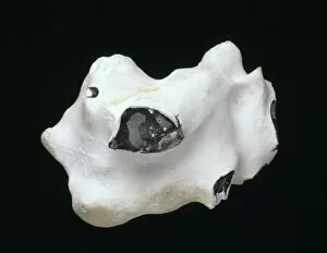
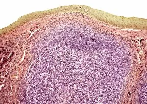
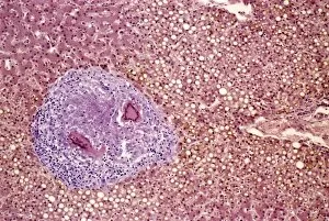
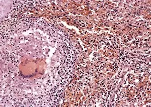
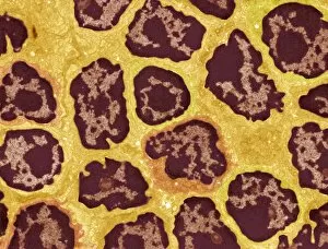
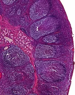
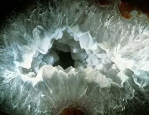
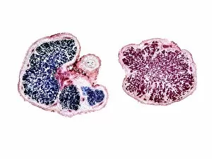
In the quaint town of Buxton, England, lies a hidden treasure known as hematite nodules. These mesmerizing formations are a sight to behold, with their deep red hues and intricate patterns. Agate nodules also grace this region, showcasing nature's artistic flair. But it is not just in Buxton where these fascinating they can be found. In far-flung corners of the world like Antarctica, they continue to captivate us. As humpback whales gracefully surface in the Southern Ocean off the Antarctic Peninsula, we catch glimpses of their majestic flukes breaking through the water's surface. Meanwhile, beneath our feet lie other remarkable discoveries waiting to be unearthed. Iron pyrite crystals shimmer on host rocks like fool's gold - a deceptive beauty that has fascinated many throughout history. And within claystone formations rests the enigmatic septarian concretion or septarian nodule - an ancient marvel formed by natural processes over millions of years. Not all nodules hold such geological wonders; some contain remnants from Earth's past. Pteridosperms preserved in ironstone nodules give us a glimpse into prehistoric plant life and its evolution over time. Yet not all nods go towards celebrating nature's splendor; some remind us of our mortality and vulnerability. Diseases like rodent ulcer and epithelioma manifest themselves on our skin as painful reminders that even our bodies can bear witness to struggle and decay. And then there are those rare treasures trapped within amber - tiny snapshots frozen in time for millennia - preserving fragments of ancient life forms forever etched within their golden embrace. Even cirrhosis of the liver finds its place among these diverse nods, reminding us that health too can falter amidst life's tapestry. From picturesque landscapes to microscopic worlds unseen by most eyes, nodules encompass both beauty and fragility alike. They serve as silent witnesses to Earth’s ever-changing story – each one holding secrets waiting to be discovered.