
Oral Cavity Collection
The oral cavity, a gateway to our body's intricate systems, holds the key to both nourishment and communication


All Professionally Made to Order for Quick Shipping
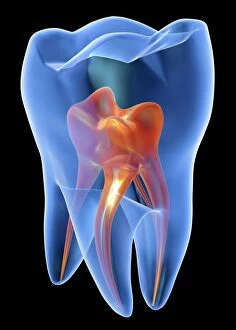
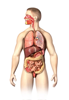
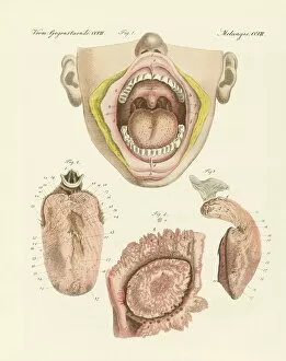
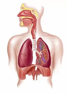
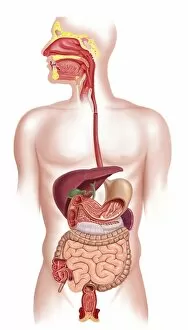
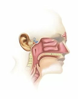
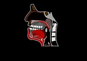
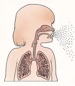
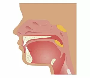
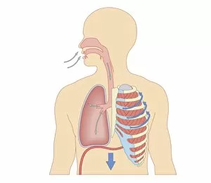
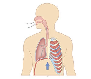
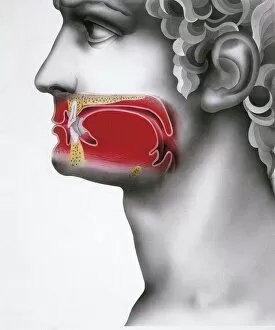
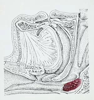
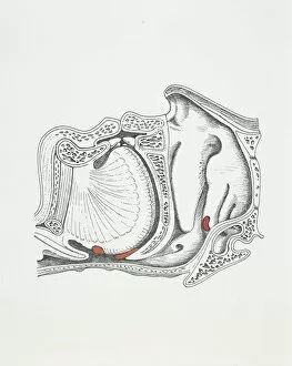
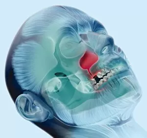
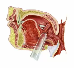
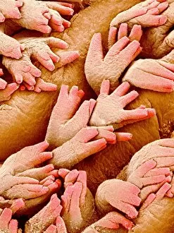
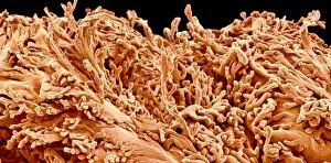
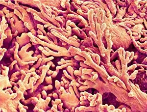
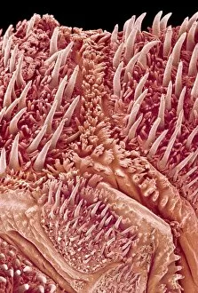
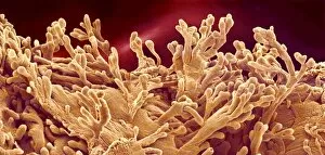
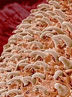
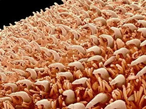
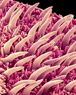
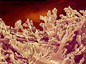
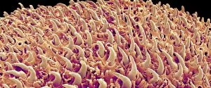
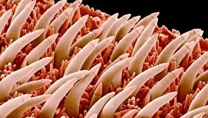
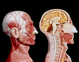
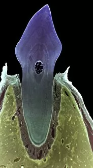
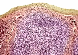
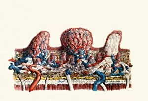
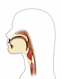
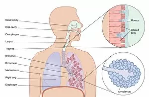
The oral cavity, a gateway to our body's intricate systems, holds the key to both nourishment and communication. Nestled within its confines are the mighty molar teeth, guardians of chewing and grinding. A cross-section biomedical illustration reveals the interconnectedness of the human digestive system as it seamlessly connects with the esophagus. This intricate network ensures that every bite we take embarks on a journey towards nourishing our bodies. While exploring this captivating realm, one cannot overlook the anatomy of the male respiratory system and internal organs, and is here that breath intertwines with life itself, sustaining us in ways beyond comprehension. Intriguingly, taste comes alive through a colored engraving showcasing its wonders. The delicate dance between flavors unfolds within this small yet powerful space - an orchestra for our senses. Delving deeper into this wondrous domain, a cutaway diagram unravels the intricacies of our respiratory system. Every inhale fuels us while every exhale releases what no longer serves us – an eternal cycle of life-giving breaths. As we navigate further into this labyrinthine landscape, a cross-section glimpse into our digestive system unveils its remarkable complexity. From stomach to intestines, each organ plays its part in breaking down food and extracting vital nutrients. Venturing even closer towards exploration lies another marvel: the anatomy of inner ear and sinuses. These hidden chambers not only aid in hearing but also maintain balance within ourselves – both physically and emotionally. Artwork depicting mouth anatomy showcases nature's masterpiece; lips forming words that connect hearts across distances vast or near. Communication finds solace here as language dances upon tongues like poetry set free. Witnessing growth from infancy onwards brings forth wonderment as seen in a cross-section illustration featuring lungs and trachea exhaling breath from mouth and nose. Each exhalation symbolizes growth - an ever-evolving testament to life's perpetual motion.