
Paediatrics Collection
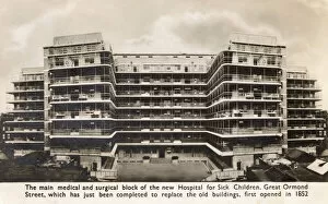
"Paediatrics: Nurturing the Future of Children's Health" In the heart of London, the New Great Ormond Street Hospital stands as a beacon of hope for little ones in need


All Professionally Made to Order for Quick Shipping
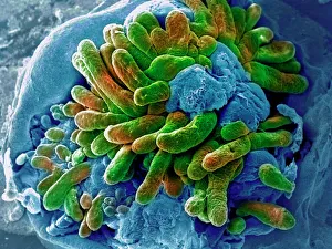
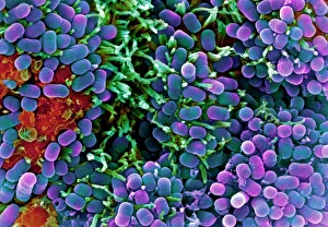
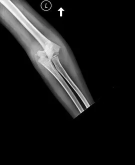
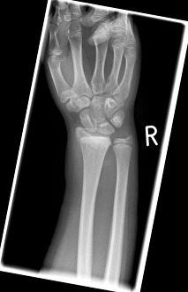
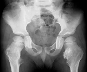
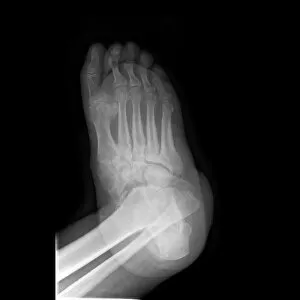
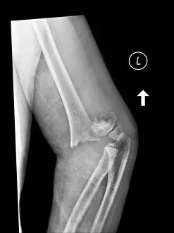
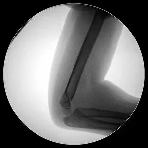
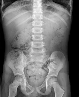
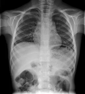
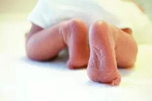
"Paediatrics: Nurturing the Future of Children's Health" In the heart of London, the New Great Ormond Street Hospital stands as a beacon of hope for little ones in need. From delicate surgeries to intricate treatments, this renowned institution is dedicated to providing exceptional care for children. Delving into the microscopic world, we witness a fascinating SEM image showcasing the intricacies of the small intestine. This captivating glimpse reminds us of the incredible complexity that paediatricians navigate daily. As parents arrive with their precious bundles, they find solace in convenient buggy parking facilities at this esteemed hospital. Ensuring ease and accessibility for families during their visits is just one way paediatrics goes above and beyond. Zooming further into another SEM image reveals E. Coli bacteria – an unwelcome intruder but a subject of study nonetheless. Paediatricians tirelessly work to combat such threats and safeguard our little ones from harm. A baby boy lies peacefully under expert care at Royal Alexandra Hospital for Children in Brighton, Sussex. Here, compassionate professionals provide comfort and healing while nurturing young lives back to health. At 1st Bellevue Scout Troop Hospital, injured scouts find solace knowing that skilled paediatricians are there to mend broken bones like those captured on X-ray C017 / 7975 – a testament to both adventure and resilience. X-ray C017 / 7561 unveils deformed hands seeking remedy within these specialised walls. With expertise honed through years of training, paediatricians strive not only for physical recovery but also emotional support during challenging times. A snapshot captures child's molars alongside an innocent tongue – symbols of growth and development within young mouths cared for by dedicated dental specialists who understand every unique smile deserves attention. Another X-ray (C017 / 7266) uncovers a broken elbow; however, it also showcases how resilient children can be when guided by skilful hands committed to restoring their mobility and joy.