
Papilla Collection
"Papilla: Unveiling the Intricate Surfaces of Nature and Life" Delve into the mesmerizing world of papilla


All Professionally Made to Order for Quick Shipping
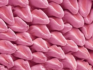
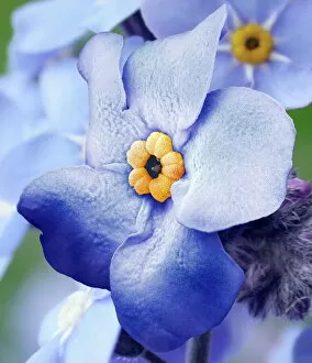
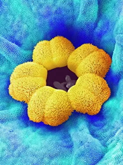
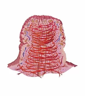
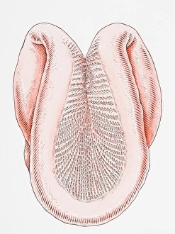
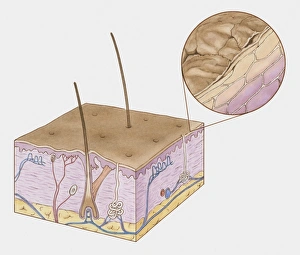
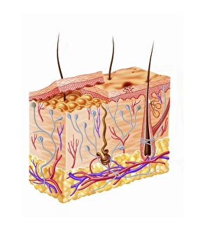
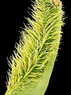
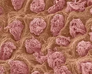
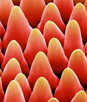
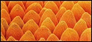
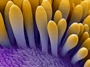
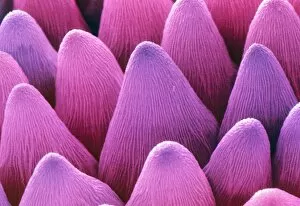
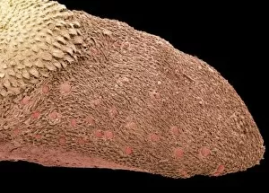
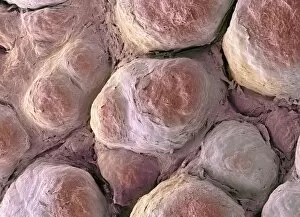
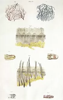
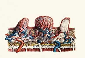
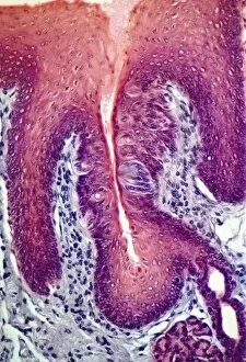
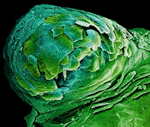
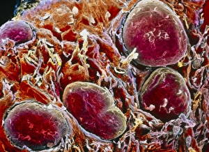
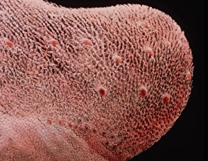
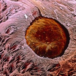
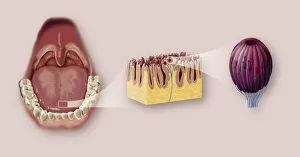
"Papilla: Unveiling the Intricate Surfaces of Nature and Life" Delve into the mesmerizing world of papilla, where intricate textures and structures reveal themselves under the lens. From the cat's tongue surface resembling a delicate rasp to forget-me-not flowers showcasing their hidden beauty, these SEM images offer a glimpse into nature's remarkable designs. Intriguingly, even our own bodies hold secrets within. Witness the erect breast nipple captured in an artwork that celebrates human form and diversity. Explore further as we journey beneath the microscope to discover cow tongues' microscopic wonders or delve into cross-sections of a normal heart, unraveling its complex anatomy. But it doesn't stop there; let us guide you through illustrations depicting the structure of human skin and hair. Marvel at how each layer intertwines seamlessly to protect and nourish us from external elements. Delight in diagrams unveiling every detail about this incredible organ that shields our inner selves. And finally, witness feline elegance as we observe cats curling their tongues like ladles to lap up liquid effortlessly. Each papilla pointing backward ensures maximum efficiency while drinking - truly a marvel of evolution. Join us on this captivating journey through papilla's diverse realms - from animal tongues to flower petals, from hearts beating with life to intricate layers protecting our very existence. Prepare for awe-inspiring revelations that will leave you appreciating nature's artistry like never before.