
Parasitism Collection
"Unseen Intruders: Exploring the World of Parasitism" Caption: In this captivating series of images, we delve into the intricate and often hidden world of parasitism


All Professionally Made to Order for Quick Shipping
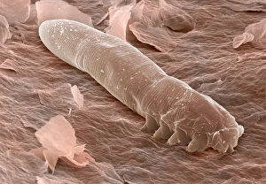
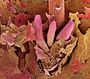
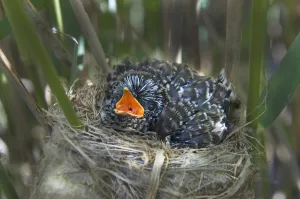
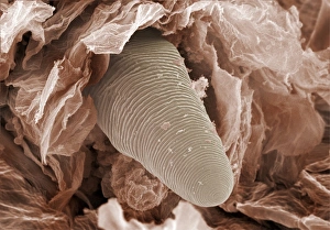
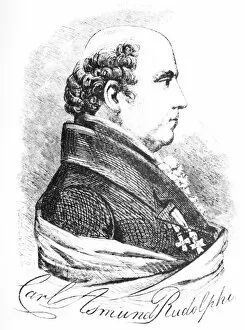
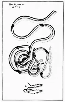
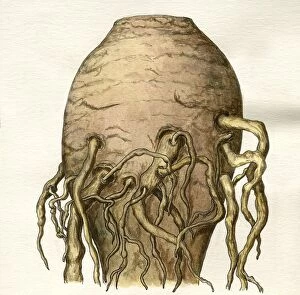
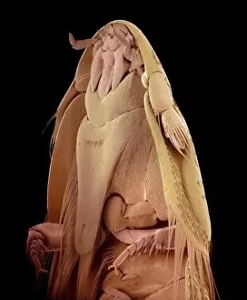
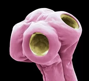
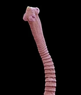
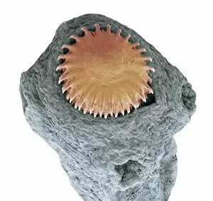
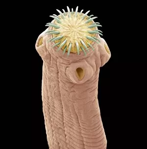
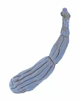
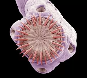
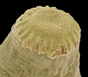
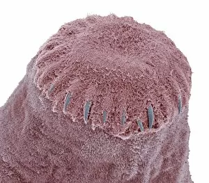
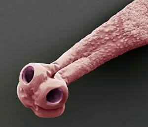
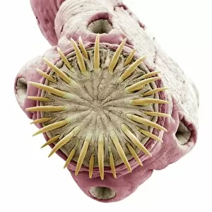
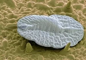
"Unseen Intruders: Exploring the World of Parasitism" Caption: In this captivating series of images, we delve into the intricate and often hidden world of parasitism. Starting with a close-up view captured by Scanning Electron Microscopy (SEM), we witness the astonishing presence of eyelash mites (Picture No. 12479415). These tiny creatures, barely visible to the naked eye, inhabit our very own eyelashes, reminding us that even within our bodies, parasites can thrive. Zooming in further with SEM technology, another image reveals the intriguing tails of these eyelash mites (Picture No. 12479414). Their peculiar structures hint at their evolutionary adaptations for survival and reproduction. Moving beyond microscopic organisms, we encounter an extraordinary example of brood parasitism involving a Cuckoo chick in a Reed Warbler nest (Acrocephalus scirpaceus) (INDO 20-06 393). Picture No. 12479506 showcases a twelve-day-old European Cuckoo chick nestled among unsuspecting Reed Warblers in Africa. This remarkable behavior is not without consequences as native species are affected by sleeping sickness due to such intrusions. The historical engraving by Karl Rudolphi portrays this phenomenon vividly; it serves as a reminder that parasitic interactions have fascinated naturalists throughout history (Picture No. 12479416). The image depicts an African scene where a twelve-day-old European Cuckoo chick thrives at the expense of its foster parents' offspring while contributing to ecological imbalances. Lastly, we witness firsthand how nature's delicate balance can be disrupted when observing a Reed Warbler diligently feeding its adopted twelve-day-old Cuckoo chick (Cuculus canorus) despite being unrelated species (Acrocephalus scirpaceus). These captivating visuals shed light on the intricacies and complexities surrounding parasitism—a phenomenon both fascinating and often unsettling.