Fraud Blocker Skip to main content

Pathology Collection

Background imagePathology Collection: Set of glass eyeballs

Set of glass eyeballs. Each shell illustrates a disease or abnormality affecting the eye. The set was hand crafted in the late 1920s by ocularists of Theodore Hamblin Ltd

Background imagePathology Collection: Sleeping sickness parasite

Sleeping sickness parasite
Sleeping sickness. Artwork of a trypanosome (Trypanosoma brucei) moving past human red blood cells in the blood. This protozoan is the cause of sleeping sickness (African trypanosomiasis)

Background imagePathology Collection: Human brain microscope slides

Human brain microscope slides. Microscope slides of stained sections of human brain. These slides are from the Harvard Brain and Tissue Resource Centre, USA

Background imagePathology Collection: Tobacco hornworm with tobacco plant

Tobacco hornworm with tobacco plant
Tobacco hornworm (Manduca sexta) with a damaged tobacco plant, artwork. This species is common throughout North America

Background imagePathology Collection: Alzheimers brain

Alzheimers brain. Computer processed vertical (coronal) slice through the brain of an Alzheimer patient (at left) compared with a normal brain (at right)

Background imagePathology Collection: Gout crystals

Gout crystals. Polarised light micrograph of uric acid crystals in the synovial fluid (a lubricating liquid in joints) from a patient with gout

Background imagePathology Collection: Dohle bodies in blood cell, micrograph

Dohle bodies in blood cell, micrograph
Dohle bodies in blood cell. Light micrograph of a neutrophil white blood cell (centre) with Dohle bodies. These are small inclusions within the cells cytoplasm

Background imagePathology Collection: Liver tissue cirrhosis, light micrograph

Liver tissue cirrhosis, light micrograph
Liver cirrhosis. Light micrograph of a section through liver tissue showing cirrhosis

Background imagePathology Collection: Louis Pasteur, French microbiologist

Louis Pasteur, French microbiologist
Louis Pasteur (1822-1895), French microbiologist and chemist. Louis Pasteur proved the germ theory of disease, which holds that germs attack the body from the outside

Background imagePathology Collection: Microscopic view of human respiratory syncytial virus

Microscopic view of human respiratory syncytial virus (RSV). RSV causes respiratory tract infection of the lungs and breathing passages

Background imagePathology Collection: Microscopic view of chlamydia

Microscopic view of chlamydia. Chlamydia is a common sexually transmitted disease (STD) caused by a bacterium

Background imagePathology Collection: Acute promyelocytic leukaemia, micrograph

Acute promyelocytic leukaemia, micrograph
Acute promyelocytic leukaemia. Light micrograph of blood cells from bone marrow in a case of acute promyelocytic leukaemia. Leukaemia is a cancer where certain blood cells form in excess

Background imagePathology Collection: Liver cirrhosis

Liver cirrhosis. Artwork taken from Sir Robert Carswells textbook Pathological Anatomy, published 1838

Background imagePathology Collection: Cystic fibrosis

Cystic fibrosis. Light micrograph of a section through a bronchus (airway) of the lungs in a case of cystic fibrosis (mucoviscidosis)

Background imagePathology Collection: Louis Pasteur, French microbiologist

Louis Pasteur, French microbiologist
Louis Pasteur (1822-1895), French microbiologist and chemist, in his laboratory. Louis Pasteur proved the germ theory of disease, which holds that germs attack the body from the outside

Background imagePathology Collection: Neutrophil cell trapping bacteria, SEM

Neutrophil cell trapping bacteria, SEM
Neutrophil cell trapping bacteria. Coloured scanning electron micrograph (SEM) of bacteria (rod-shaped) being trapped by a neutrophil cell

Background imagePathology Collection: Osteoporotic bone

Osteoporotic bone

Background imagePathology Collection: Klebsiella pneumoniae clothed as devil cartoon character design concept

Klebsiella pneumoniae clothed as devil cartoon character design concept Klebsiella pneumoniae clothed as devil cartoon character design concept. Vector illustration

Background imagePathology Collection: Rudolf Virchow (1821-1902) was a German pathologist and founder of cell pathology

Rudolf Virchow (1821-1902) was a German pathologist and founder of cell pathology. He later devoted himself to anthropology and archaeology and worked with Schliemann on the excavations at Troy

Background imagePathology Collection: Rudolph Virchow (1821-1902) German pathologist and founder of cell pathology

Rudolph Virchow (1821-1902) German pathologist and founder of cell pathology. He later devoted himself to anthropology and archaeology and worked with Schliemann on the excavations at Troy

Background imagePathology Collection: Ambroise Pare, c. 1510-1590, a French barber surgeon

Ambroise Pare, c. 1510-1590, a French barber surgeon

Background imagePathology Collection: Ambroise Pare, c. 1510-1590, a French barber surgeon

Ambroise Pare, c. 1510-1590, a French barber surgeon

Background imagePathology Collection: Sir Astley Paston Cooper, 1st Baronet

Sir Astley Paston Cooper, 1st Baronet, 1768-1841, an English surgeon and anatomist, Sir Astley Paston Cooper, 1768-1841

Background imagePathology Collection: Diagram showing different skin conditions

Diagram showing different skin conditions Diagram showing different skin conditions illustration

Background imagePathology Collection: Mycobacterium cartoon character design style with board

Mycobacterium cartoon character design style with board Mycobacterium cartoon character design style with board. Vector illustration

Background imagePathology Collection: Complications of gastric ulcer. Bleeding and perforation

Complications of gastric ulcer. Bleeding and perforation

Background imagePathology Collection: Information poster of human heart diagram illustration

Information poster of human heart diagram illustration

Background imagePathology Collection: Colon cancer tissue section under the microscope 100x

Colon cancer tissue section under the microscope 100x

Background imagePathology Collection: Zika virus infographic: prevention, symptoms and treatment

Zika virus infographic: prevention, symptoms and treatment

Background imagePathology Collection: Dr. Karl Landsteiner

Dr. Karl Landsteiner
(Original Caption) 11/1/30-New York: Closeup of Dr. Karl Landsteiner, famous bacteriologist and pathologist, who has just been announced as the winner of the 1930 Nobel prize for medicine. Dr

Background imagePathology Collection: Jonas Salk and Luc Montagnier Hug

Jonas Salk and Luc Montagnier Hug
AIDS researcher Luc Montagnier hugs Jonas Salk, developer of the polio vaccine, after they were both presented medical research awards by the Albert and Mary Lasker Foundation

Background imagePathology Collection: Rudolph Virchow Looking Through Magnifying Glass

Rudolph Virchow Looking Through Magnifying Glass
(Original Caption) Rudolph Virchow. Photograph

Background imagePathology Collection: Pathological laboratory - COMFORT, between c1915 and c1920. Creator: Bain News Service

Pathological laboratory - COMFORT, between c1915 and c1920. Creator: Bain News Service
Pathological laboratory - COMFORT, between c1915 and c1920

Background imagePathology Collection: Illustration of Edward Jenner Vaccinating James Phipps

Illustration of Edward Jenner Vaccinating James Phipps
(Original Caption) Edward Jenner vaccinating James Phipps, a boy of eight, May 14, 1796. Pus from the hand of Sarah Nelmes, a dairy maid, was used. Artist unknown. Undated illustration

Background imagePathology Collection: A Flea

A Flea
(Original Caption) Dog's Nitemare. Schenectady, New York: No wonder Fido scratches, with creatures like this crawling on him

Background imagePathology Collection: Jonas Salk Holding Bottles of Polio Vaccine

Jonas Salk Holding Bottles of Polio Vaccine
Dr. Jonas Salk displays his polio vaccine which he developed in a University of Pittsburgh laboratory

Background imagePathology Collection: Jonas Salk

Jonas Salk
(Original Caption) 11/2/1956- Dr. Jonas Salk, director, Virus Research Laboratory, University of Pittsburgh, PA, who was named winner yesterday (Nov)

Background imagePathology Collection: George Papanicolaou Examining a Slide

George Papanicolaou Examining a Slide
(Original Caption) 5/13/1958-New York, NY: Dr. George N. Papanicolaou examines a slide here. "Dr

Background imagePathology Collection: James Paget

James Paget
3733770 James Paget.; (add.info.: Photographic portrait of James Paget (1814-1899) an English surgeon and pathologist, best remembered for naming Paget's Disease

Background imagePathology Collection: Measles

Measles
5855402 Measles; (add.info.: Measles, Rubeola vulgaris and R. nigra. Handcoloured copperplate stipple engraving by John Pass from John Wilkes "Encyclopedia Londinensis, " J)

Background imagePathology Collection: Purple spots and rose rash

Purple spots and rose rash
5855400 Purple spots and rose rash; (add.info.: Skin diseases: Purple spots, Purpura simplex and P. haemorrhagica and rose rash, Exanthesis roseola)

Background imagePathology Collection: Psoriasis

Psoriasis
5855396 Psoriasis; (add.info.: Skin diseases: Prurigo nodularis and Psoriasis varieties)

Background imagePathology Collection: Scalp and skin diseases

Scalp and skin diseases
5855397 Scalp and skin diseases; (add.info.: Scalp diseases Porrigo crustacea and P. furfuracea, and skin diseases Ecthyma vulgaris and E)

Background imagePathology Collection: Sycosis

Sycosis
5855392 Sycosis; (add.info.: Sycosis barbae and Sycosis capilli, chronic inflammation of the hair follicles, especially of the beard and head)

Background imagePathology Collection: Sir James Paget, Victorian period

Sir James Paget, Victorian period

Background imagePathology Collection: Professor Rudolf Virchow (engraving)

Professor Rudolf Virchow (engraving)
1038752 Professor Rudolf Virchow (engraving) by Wirgman, Theodore Blake (1848-1925) (after); Private Collection; (add.info.: Professor Rudolf Virchow)

Background imagePathology Collection: Queen Elizabeth Hospital, Sheriff Hill, Gateshead, England. 19th February 1986

Queen Elizabeth Hospital, Sheriff Hill, Gateshead, England. 19th February 1986. Pathology Lab

Background imagePathology Collection: Representation of a trepanation in Ancient Egypt

Representation of a trepanation in Ancient Egypt. Painting

Background imagePathology Collection: Comparison of healthy gums versus gingivitis

Comparison of healthy gums versus gingivitis

Background imagePathology Collection: Histopathology and pathophysiology of diabetic food ulcers

Histopathology and pathophysiology of diabetic food ulcers

Background imagePathology Collection: Santiago Ramon and Cajal (1852-1934) Spanish histologist, physician and pathologist

Santiago Ramon and Cajal (1852-1934) Spanish histologist, physician and pathologist
Santiago Ramon and Cajal (1852-1934).. Spanish histologist, physician and pathologist. He made important discoveries such as laws governing the morphology and connections of nerve cells in the brain

Background imagePathology Collection: Jean Martin Charcot demonstrating hypnosis, 1879

Jean Martin Charcot demonstrating hypnosis, 1879. Charcot (1825-1893), French neurologist and pathologist, demonstrating the production of hypnosis using beam of light from a magic lantern

Background imagePathology Collection: Enfermedades de los ojos (Eye diseases). Engraving

Enfermedades de los ojos (Eye diseases). Engraving illustrating major eye diseases: stye, conjunctivitis, squint, pterygium, keratitis. Engraving

Background imagePathology Collection: Cross-section diagram of a cancerous tumour including calcium deposits

Cross-section diagram of a cancerous tumour including calcium deposits, blood vessels, tumour outgrowth, epithelial layer, ulcerated area, bleeding, nerve fibres, dead tissue and a lymph vessel

Background imagePathology Collection: Scanning electron micrograph of HIV particles infecting a human H9 T cell

Scanning electron micrograph of HIV particles infecting a human H9 T cell

Background imagePathology Collection: Coxiella burnetii, the bacteria that causes Q Fever

Coxiella burnetii, the bacteria that causes Q Fever. A dry fracture of a Vero cell exposing the contents of a vacuole where Coxiella burnetii are busy growing

Background imagePathology Collection: Stylized rabies virus particles

Stylized rabies virus particles, the cause of the viral neuroinvasive disease acute encephalitis

Background imagePathology Collection: Anatomy of superficial (surface) lymphatics

Anatomy of superficial (surface) lymphatics

Background imagePathology Collection: Microscopic view of a leukemia cell

Microscopic view of a leukemia cell

Background imagePathology Collection: Microscopic view of Neisseria gonorrhoeae

Microscopic view of Neisseria gonorrhoeae

Background imagePathology Collection: Conceptual image of the Hepatitis C virus

Conceptual image of the Hepatitis C virus. Hepatitis C virus is a small, enveloped, positive-sense single-stranded RNA virus of the family Flaviviridae

Background imagePathology Collection: Trachea cross-section with lungs showing normal and asthmatic bronchiole

Trachea cross-section with lungs showing normal and asthmatic bronchiole
Trachea cross-section with lungs, showing comparison of normal and asthmatic bronchiole

Background imagePathology Collection: Good and bad cholesterol found in blood stream

Good and bad cholesterol found in blood stream

Background imagePathology Collection: Large eroded plaque on the leg with areas of crust

Large eroded plaque on the leg with areas of crust
Metastatic Melanoma. Large eroded plaque on the leg with areas of crust. Scattered eroded dermal nodules are also present

Background imagePathology Collection: Melanoma on a patients skin

Melanoma on a patients skin

Background imagePathology Collection: Acute coronary syndrome - microvascular obstruction

Acute coronary syndrome - microvascular obstruction


All products are expertly crafted, using premium materials, tailored to your specifications and promptly shipped


EDITORS COMMENTS

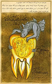
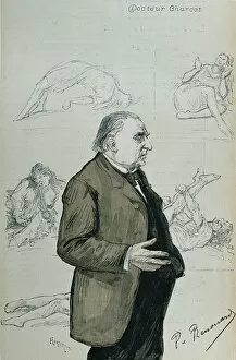
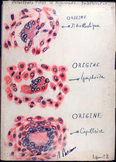
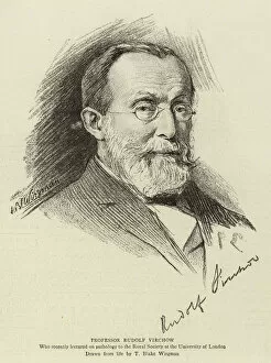
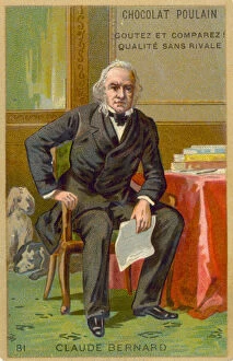
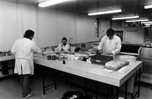
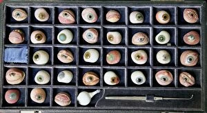
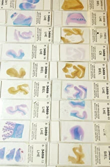
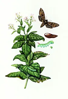
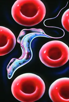
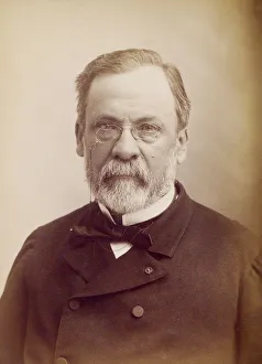
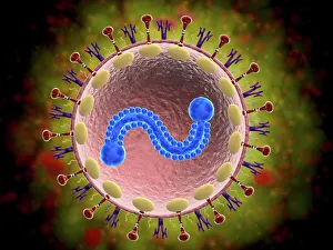
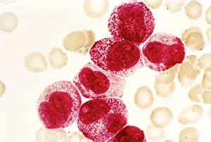
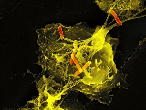
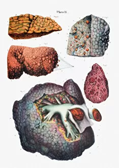
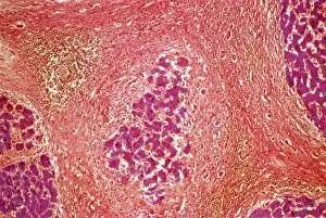
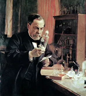
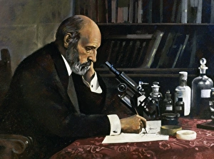
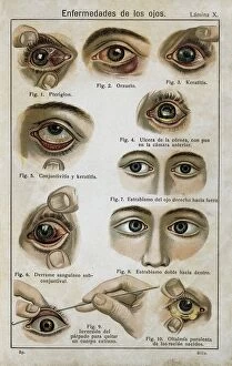
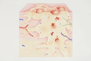
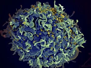
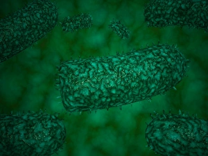
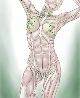
"Exploring the Intricate World of Pathology: Unveiling the Mysteries Within" Delving into the realm of pathology, we encounter a fascinating array of scientific wonders. From a set of glass eyeballs that have witnessed countless medical marvels to human brain microscope slides revealing intricate neural networks, each specimen holds secrets waiting to be unraveled. Intriguingly, a tobacco hornworm intertwined with its tobacco plant counterpart reminds us of the intricate relationship between organisms and their environment. Similarly, an Alzheimer's brain reveals the devastating effects this disease has on our most precious organ. As we journey through history, we encounter Louis Pasteur, the renowned French microbiologist whose groundbreaking discoveries paved the way for modern medicine. His contributions continue to inspire generations in their pursuit of understanding diseases at their core. Peering through microscopes, we witness captivating views such as that of a microscopic respiratory syncytial virus invading its host or Dohle bodies within blood cells – tiny anomalies holding vital diagnostic clues. The microscopic view of chlamydia serves as a reminder that even seemingly inconspicuous pathogens can wreak havoc on our health. Examining acute promyelocytic leukemia under magnification brings awareness to the battle fought by patients and healthcare professionals alike against this aggressive form of cancer. Meanwhile, glimpsing at sleeping sickness parasites highlights how these minuscule invaders disrupt lives in regions plagued by poverty and limited resources. The sight of liver cirrhosis evokes contemplation about lifestyle choices and their consequences on one's vital organs. In contrast, light micrographs showcasing liver tissue cirrhosis provide insight into cellular changes occurring within this complex disease process. Pathology is not merely confined to textbooks; it is an ever-evolving field where scientists tirelessly strive to decipher nature's enigmatic puzzles. Each slide represents more than just pixels; they hold stories waiting to be told – tales that shed light on diseases afflicting humanity and offer hope for better treatments.

© Copyright Media Storehouse, All Rights Reserved 2003 - 2025
Facebook     Pinterest     100% Payment Secure     Reviews IO