
Pelvic Collection
"The Pelvic: A Fascinating Intersection of Art, Anatomy, and Health" The pelvic region has captivated artists and scientists alike throughout history


All Professionally Made to Order for Quick Shipping
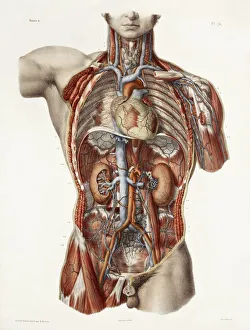
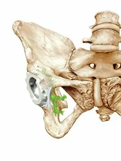
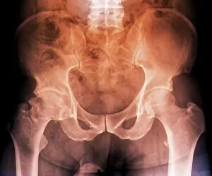
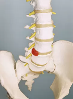
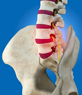
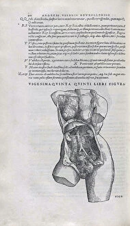
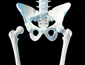
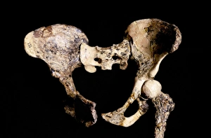
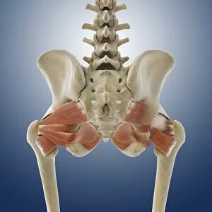
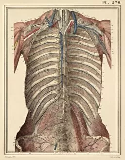
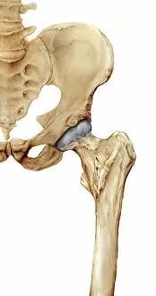
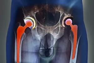
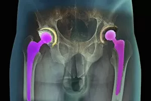
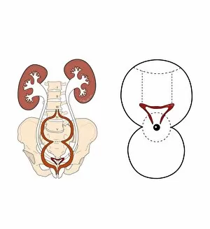
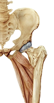
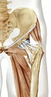
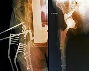
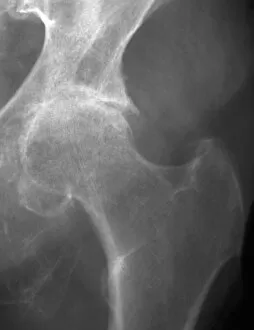
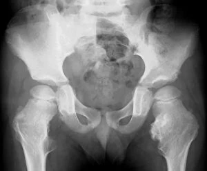
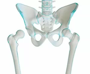
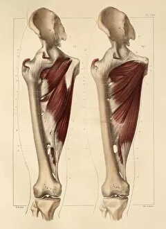
"The Pelvic: A Fascinating Intersection of Art, Anatomy, and Health" The pelvic region has captivated artists and scientists alike throughout history. From ancient historical artwork depicting the cardiovascular system's connection to the pelvis to modern depictions of hip replacement surgeries, this area holds immense significance. One common ailment that affects the pelvic region is osteoarthritis of the hip. X-ray images reveal its impact on the bones, prompting advancements in treatments like hip replacements. These procedures have revolutionized healthcare by restoring mobility and alleviating pain for countless individuals. Another condition associated with the pelvis is a slipped disc. Artists have beautifully portrayed this issue through intricate artworks showcasing slipped intervertebral discs. Such visuals help medical professionals understand and treat this painful condition effectively. Delving deeper into anatomy, X-rays provide detailed views of hip bones from various angles. The complexity of these structures becomes evident as we explore their connections to muscles such as Levator ani - an engraving displaying a side view of the pelvis highlights this relationship. The wonderment surrounding life begins within the womb, where diagrams depict foetuses developing within their mothers' pelvises. This glimpse into human reproduction showcases how essential a healthy pelvic structure is for nurturing new life. Artists have also contributed to our understanding of skeletal anatomy by creating stunning artwork portraying human skeletons in intricate detail. By studying these pieces, we gain insights into how different parts interact within the body's framework. Amidst all these scientific explorations lies an unexpected surprise – a blacktip reef shark. Though not directly related to human anatomy or health concerns involving the pelvic region, it serves as a reminder that nature's wonders extend beyond our own bodies. Returning back to humans, 1831 artwork focuses on another crucial aspect: pelvic-femoral muscles. Understanding their functions aids in comprehending movement patterns and potential issues that may arise when they are compromised. Lastly, artistic representations shed light on the complexity of hip joint bones and their anatomy.