
Photoreceptor Collection
Photoreceptors are the remarkable cells that enable us to perceive and interpret the world through our eyes


All Professionally Made to Order for Quick Shipping
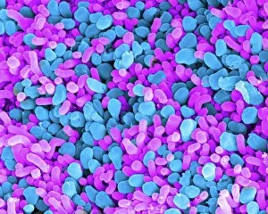
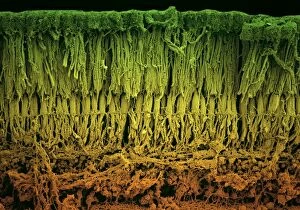
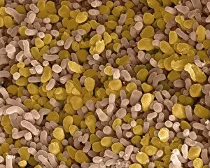
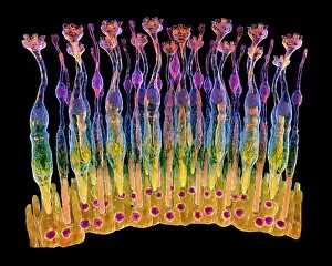
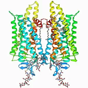
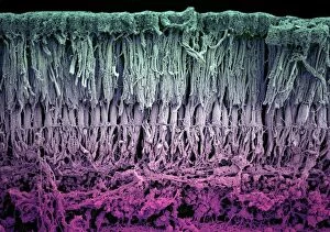
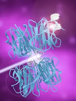
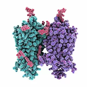
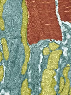
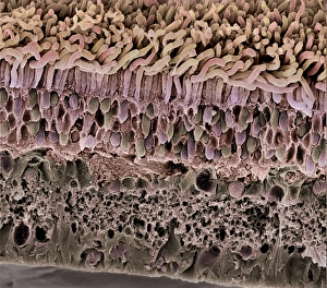
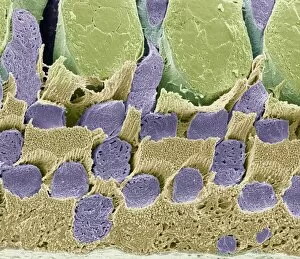
Photoreceptors are the remarkable cells that enable us to perceive and interpret the world through our eyes. These specialized cells, known as rod and cone cells, play a crucial role in our visual system. In this captivating image (SEM C014 / 4866), we get a close-up view of these photoreceptor cells. The intricate structure of each cell is beautifully captured, showcasing their unique features. Rod cells, which are responsible for vision in dim light conditions, can be identified by their elongated shape and abundant presence throughout the retina. Moving on to another stunning picture (Picture No. 12479375), we delve deeper into the world of photoreceptors with a focus on rhodopsin protein molecules. Rhodopsin is an essential component found within rod cells that aids in capturing light photons and initiating the process of vision. Continuing our exploration, we encounter yet another mesmerizing image (SEM C014 / 4864) depicting both rod and cone cells side by side (Picture No. 11675579). Cone cells are responsible for color vision and function best under bright lighting conditions. Their distinctive shape sets them apart from rods, appearing more conical or tapered. As we zoom out to observe the entire eye retina (F008 / 0713), it becomes evident how densely packed these photoreceptor cells are within this delicate tissue layer. The complexity of this network is further highlighted in subsequent images (F008 / 0719, F008 / 0714). The eye retina serves as nature's canvas where millions of photoreceptors work harmoniously to transmit visual information to our brain for processing and interpretation. Each individual cell contributes its part towards creating a vivid perception of our surroundings. This collection of images showcases not only the beauty but also the incredible functionality behind these tiny yet powerful photoreceptor units present within our eyes' retinas (F008 / 0712 - F008 / 0717).