
Physiological Collection
"Exploring the Intricacies of Physiology: From Airpumps to Blood Cells" Delving into the world of physiology


All Professionally Made to Order for Quick Shipping
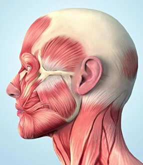
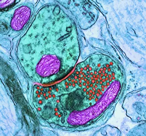
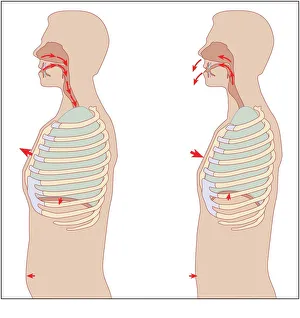
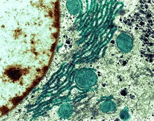
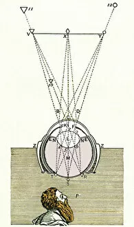
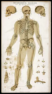
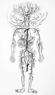
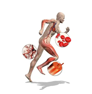
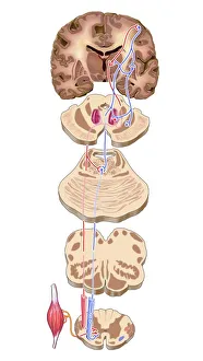
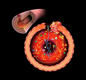
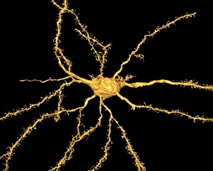
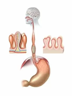
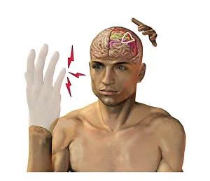
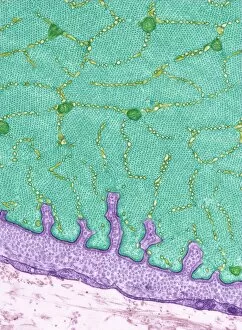
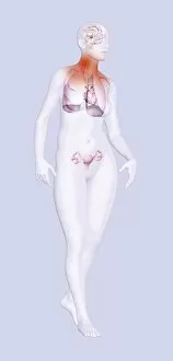
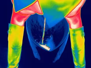
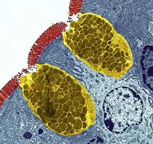
"Exploring the Intricacies of Physiology: From Airpumps to Blood Cells" Delving into the world of physiology, we uncover a fascinating array of scientific discoveries and visual wonders. Joseph Wright's masterpiece, "The Airpump, " captures the essence of early physiological experiments, showcasing humanity's thirst for knowledge. Intricate synapse nerve junctions depicted through Transmission Electron Microscopy (TEM) reveal the intricate communication network within our bodies. These microscopic connections enable seamless transmission of signals between neurons, shaping our thoughts and actions. Moving upwards to the head, we encounter an astonishing view of the muscular system. The complexity and precision required for even simple movements become apparent as ligaments intertwine with bones in perfect harmony. Venturing deeper into female physiology, a Scanning Electron Microscope (SEM) reveals the uterus lining during menstruation. This glimpse into nature's cyclical process sheds light on one aspect of womanhood that has both fascinated and perplexed scientists throughout history. Stepping away from biology but still within the realm of physiology, lie detector tests emerge as tools to decipher truth from deception. By monitoring physiological responses such as heart rate and perspiration levels, these tests attempt to unravel hidden truths buried within our bodies. Returning to cellular structures through TEM imaging once again unveils rough endoplasmic reticulum - a vital component responsible for protein synthesis in cells. Its intricate web-like structure showcases nature's elegance at work. Tracing back centuries ago brings us to Descartes' optics theory from the 17th century - an exploration into how vision is perceived by our eyes and interpreted by our brains. This groundbreaking theory laid foundations for understanding human perception today. Transitioning towards physicality itself leads us to skeletons intertwined with ligaments - providing structural support while allowing flexibility in movement. These silent heroes ensure smooth locomotion throughout life's journey.