
Phytoplankton Collection
Phytoplankton, the microscopic wonders of our aquatic world, never cease to amaze us with their intricate beauty and vital role in our ecosystem


All Professionally Made to Order for Quick Shipping
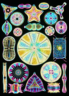
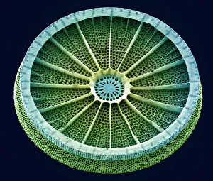
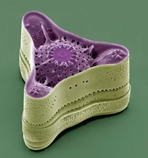
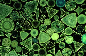
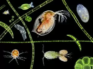
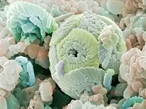
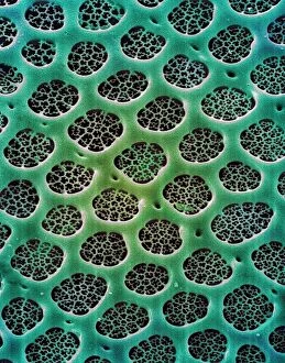
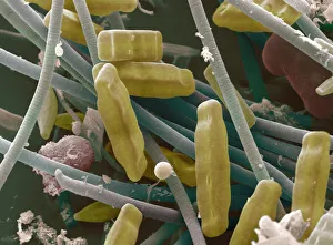
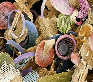
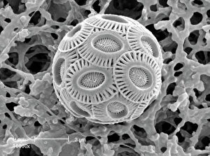
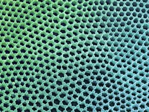
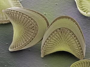
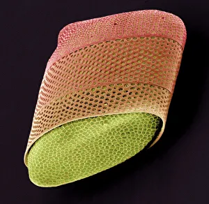
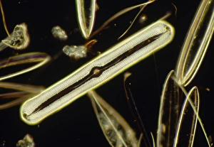
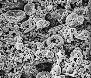
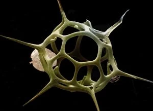
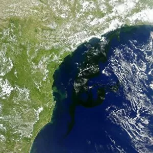
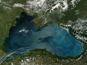
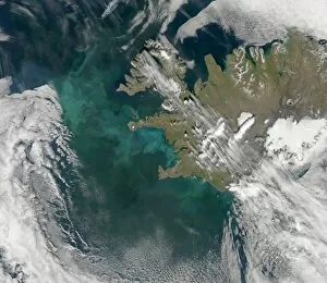
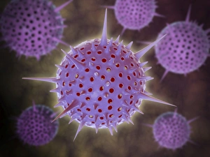
Phytoplankton, the microscopic wonders of our aquatic world, never cease to amaze us with their intricate beauty and vital role in our ecosystem. From the mesmerizing art of diatom algae captured by Ernst Haeckel to the stunning images of calcareous phytoplankton under scanning electron microscopy (SEM), these tiny organisms hold a wealth of secrets waiting to be explored. Diatoms, one of the most abundant types of phytoplankton, reveal themselves through SEM as delicate structures adorned with intricate patterns. Their cell walls, meticulously observed at high magnification, showcase an astonishing level of detail that rivals any masterpiece created by human hands. These diatoms are not only found in marine environments but also thrive in freshwater habitats like ponds where they contribute to the vibrant tapestry of pond life. The significance extends beyond their aesthetic appeal; they play a crucial role in global carbon cycling and oxygen production. Emiliana huxleyi, a coccolithophore species among them, forms beautiful calcite plates known as coccoliths that can be seen under SEM. These tiny fossils provide valuable insights into Earth's history and evolution. As we delve deeper into understanding these remarkable organisms through advanced imaging techniques such as SEM, we uncover more about their ecological importance and potential applications for various industries including biotechnology and biofuel production. The study and preservation of fossilized diatoms further aid scientists in deciphering past environmental conditions while shedding light on future climate change scenarios. In this captivating journey through microcosms unseen by the naked eye lies an appreciation for nature's intricacy and resilience. Phytoplankton remind us that even within seemingly insignificant beings lie profound contributions to sustaining life on our planet – a reminder worth cherishing as we strive towards protecting our fragile ecosystems for generations to come.