
Platelet Collection
Platelets, the unsung heroes of our bloodstream, play a vital role in the intricate dance of blood coagulation cascade


All Professionally Made to Order for Quick Shipping
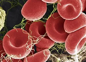
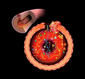
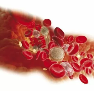
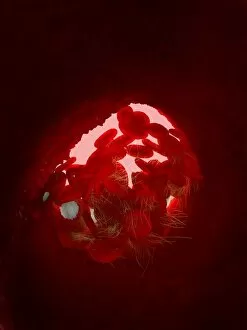
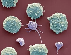
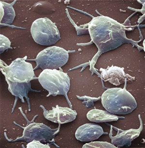
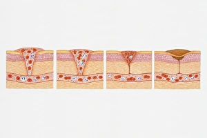
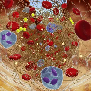
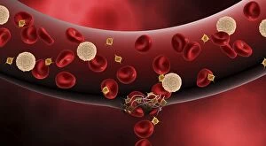
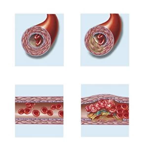
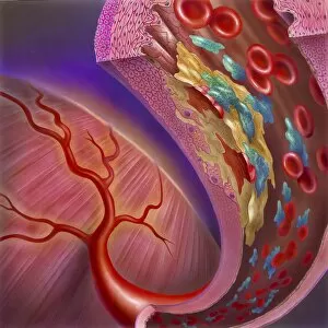
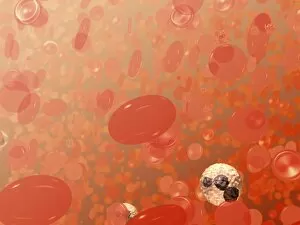
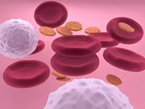
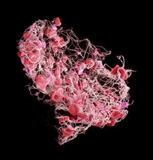
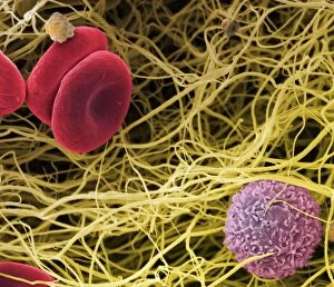
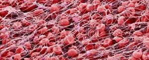
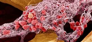
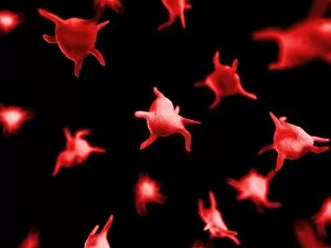
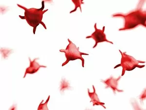
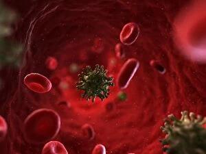
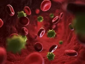
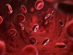
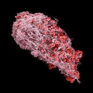
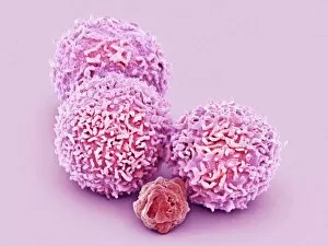
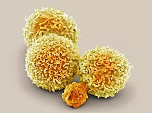
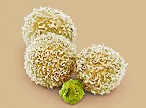
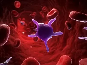
Platelets, the unsung heroes of our bloodstream, play a vital role in the intricate dance of blood coagulation cascade. Like tiny warriors they can always ready to spring into action when injury strikes. In artwork C016 / 9873, we witness the mesmerizing beauty of platelets forming a blood clot. This SEM image (C016 / 9747) captures their delicate structure as they intertwine and create a protective barrier against further bleeding. A diagram showcasing the bloodstream inside a vein reveals the harmonious collaboration between red and white blood cells alongside platelets. Together, they ensure our body's defense mechanism remains intact. SEM images of platelets demonstrate their unique appearance and highlight their importance in maintaining our health (C016). In another conceptual image featuring red blood cells and white blood cells, we see how platelets seamlessly integrate with other components to preserve equilibrium within our circulatory system. The formation of a blood clot is depicted through captivating artwork (C016 / 4619), while SEM images (C016 / 3099 & C016 / 3098) showcase the close relationship between white blood cells and platelets during this process. Illustrations depicting cross sections reveal how wounds below the skin trigger long fibrin threads that trap red blood cells while yellow-hued platelets come together to form an essential plug. In yet another biomedical illustration, damaged blood vessels constrict as activated platelets adhere to their walls - an extraordinary sight that showcases nature's remarkable ability to heal itself. Platelets may be small in size but are mighty in function. They tirelessly work behind-the-scenes ensuring our bodies can effectively respond to injuries by forming life-saving clots. Let us appreciate these incredible cellular superheroes for safeguarding our well-being every day.