
Pollen Grain Collection
"Exploring the Intricate World of Pollen Grains: A Captivating SEM Journey" Step into the fascinating realm of pollen grains, where beauty and science intertwine


All Professionally Made to Order for Quick Shipping
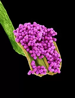
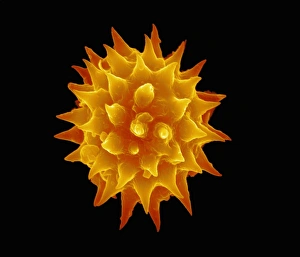
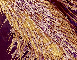
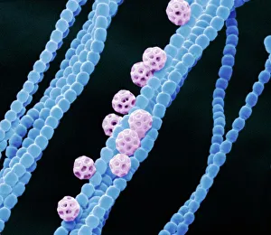
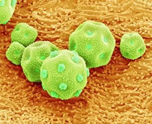
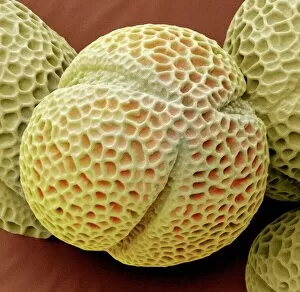
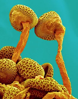
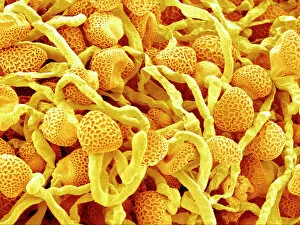
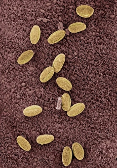
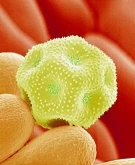
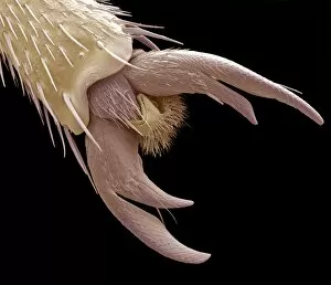
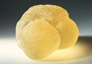
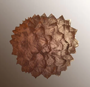
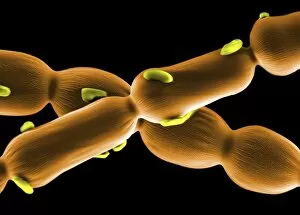
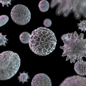
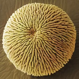
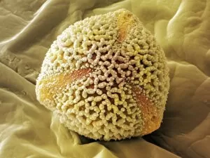
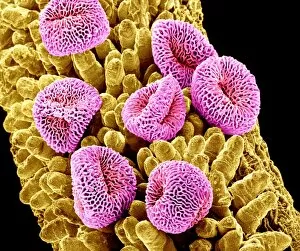
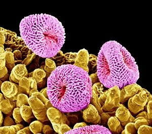
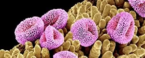
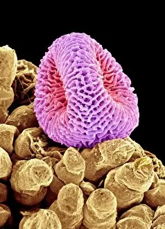
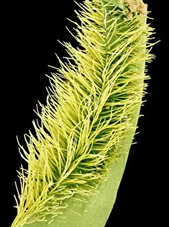
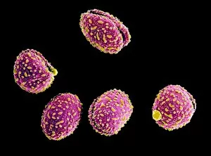
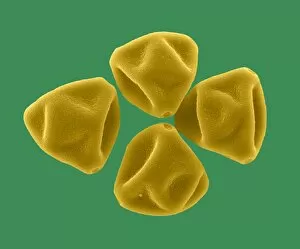
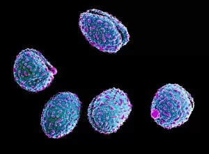
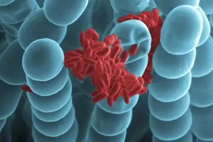
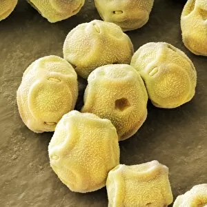
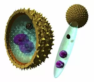
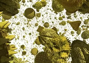
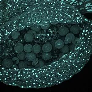
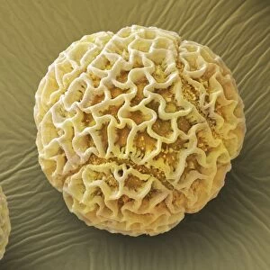
"Exploring the Intricate World of Pollen Grains: A Captivating SEM Journey" Step into the fascinating realm of pollen grains, where beauty and science intertwine. Through scanning electron microscopy (SEM), we unveil the hidden wonders of various pollen types, each with its unique structure and purpose. Let's begin our journey with a close-up view of Geranium anther pollen under SEM. The intricate patterns and delicate features reveal nature's meticulous craftsmanship at play. Moving on to Dahlia flower pollen, SEM reveals stunningly sculpted grains that resemble tiny works of art. Their textured surfaces hold secrets waiting to be unraveled. Next up is Passion flower pollen, captured in extraordinary detail by SEM. Its distinct shape and surface ornamentation showcase nature's diversity in design. Shifting our focus to Pine pollen grains through a light micrograph, we witness their spherical forms adorned with spiky projections – a testament to their adaptation for wind dispersal. Returning to SEM imagery, Geranium pollen takes center stage once more. Zooming in reveals its intricately patterned exine layer, serving as protection during pollination journeys. A captivating sight awaits us as we observe Pollen on a bee leg through SEM imaging. These tiny particles cling onto the fuzzy surface like precious cargo carried from one bloom to another – essential for plant reproduction. Diving deeper into microscopic wonders, Cyanobacteria come into view under SEM. While not traditional plant pollens, they too contribute significantly to Earth's ecosystems and deserve admiration for their role in nitrogen fixation. Chickweed pollen grains take center stage next - meticulously detailed structures resembling miniature spheres adorned with fine ridges that hint at their evolutionary adaptations for successful pollination strategies. Hellebore pollen mesmerizes us next as it showcases its unique characteristics under SEM scrutiny - intriguing shapes coupled with finely etched textures make this grain stand out among others in the botanical world. Pollen tubes steal the spotlight as SEM reveals their intricate network within lily pollen.