
Proteins Collection
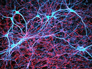
Proteins: The Building Blocks of Life From the intricate network of nerve and glial cells to the mesmerizing patterns seen under a light micrograph


All Professionally Made to Order for Quick Shipping
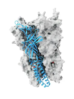
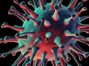
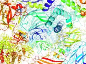
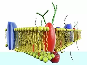
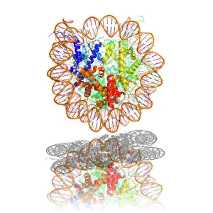
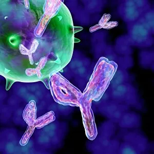
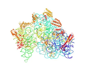
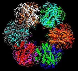
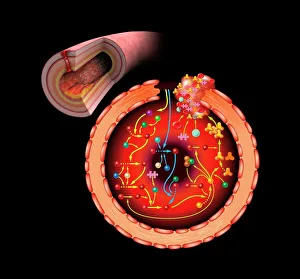
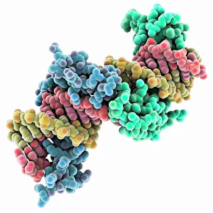
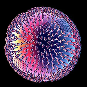
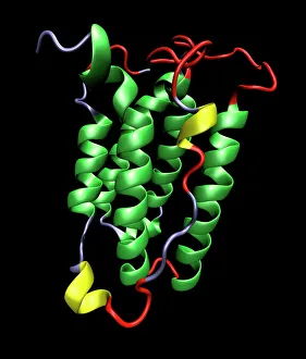
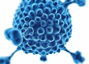
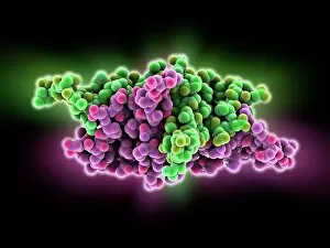
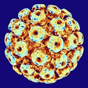
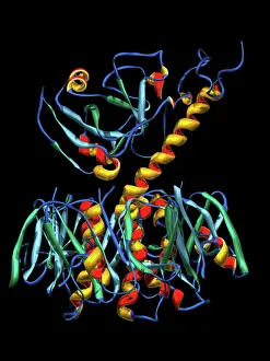
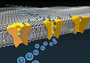
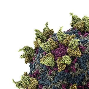
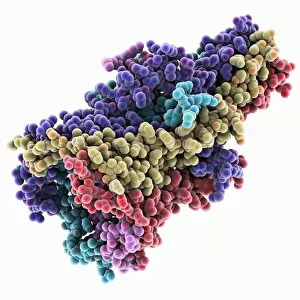
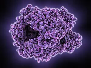
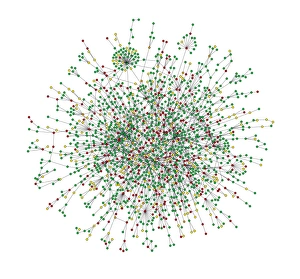
Proteins: The Building Blocks of Life From the intricate network of nerve and glial cells to the mesmerizing patterns seen under a light micrograph, proteins play an essential role in every aspect of our existence, and are like the conductors of our body's symphony, orchestrating vital processes that keep us alive and functioning. Take, for example, an anaesthetic inhibiting an ion channel C015 / 6718. Proteins act as gatekeepers, controlling what enters or exits our cells. In this case, they regulate the flow of ions necessary for transmitting nerve signals and maintaining proper cell function. But proteins don't just govern our internal workings; they also interact with external threats such as the avian flu virus. These microscopic invaders hijack host cells using their own protein machinery to replicate themselves. Understanding these interactions is crucial in developing effective treatments against viral infections. While some proteins protect us from harm, others contribute to overall well-being through a balanced diet. Our bodies require various types found in different foods to ensure optimal health and nutrition. The secondary structure is truly a work of art—a complex folding pattern that determines their shape and function. Artists have captured this beauty through stunning artwork showcasing these intricate molecular structures. One such structure is the nucleosome molecule—an elegant arrangement where DNA wraps around protein spools called histones—forming compact units within chromosomes. This organization allows efficient storage and retrieval of genetic information during cell division or gene expression. Antibodies are another remarkable class depicted in captivating artwork. These specialized molecules recognize foreign substances like bacteria or viruses and neutralize them by binding tightly to specific targets on their surface—an extraordinary defense mechanism employed by our immune system. Speaking of bacteria, their ribosomes serve as factories producing new proteins based on instructions encoded in DNA—the blueprint for life itself. Understanding bacterial ribosomes has led to groundbreaking discoveries in antibiotic development, combating infectious diseases that threaten human health.