Fraud Blocker Skip to main content

Proteins Collection (page 5)

Proteins: The Building Blocks of Life From the intricate network of nerve and glial cells to the mesmerizing patterns seen under a light micrograph

Background imageProteins Collection: Nucleus and endoplasmic reticulum F006 / 9196

Nucleus and endoplasmic reticulum F006 / 9196
Computer artwork showing part of a human or eukaryotic cell. In the middle the nucleus which has a membrane with nuclear pores. Inside the nucleus is the DNA
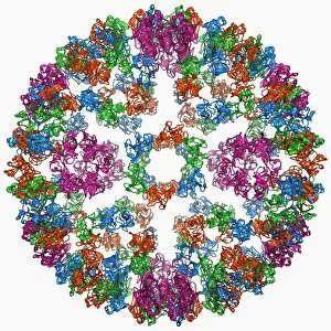
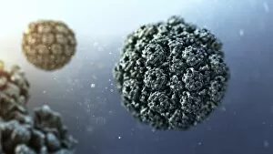
Background imageProteins Collection: Tobacco necrosis virus capsid

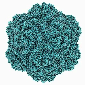
Tobacco necrosis virus capsid, molecular model. This plant virus infects a wide rage of plants, including the tobacco plant for which it is named. The virus causes tissue death (necrosis)

Background imageProteins Collection: TATA box-binding protein complex C017 / 7083

TATA box-binding protein complex C017 / 7083
TATA box-binding protein complex. Molecular model showing a TATA box-binding protein (TBP, green) complexed with a strand of DNA (deoxyribonucleic acid, yellow) and transcription factor IIB

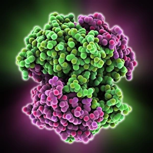
Background imageProteins Collection: Chaperonin protein F006 / 9690

Chaperonin protein F006 / 9690
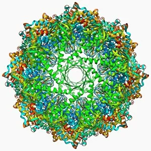
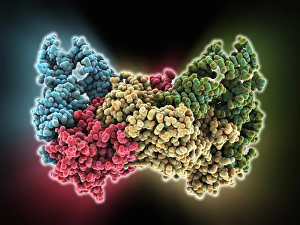
Chaperonin protein, molecular mode. Chaperonins are proteins that provide favourable conditions for the correct folding of other proteins

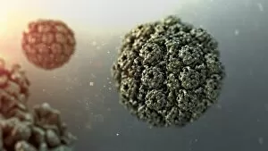
Background imageProteins Collection: Grapevine fanleaf virus capsid

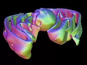
Grapevine fanleaf virus capsid, molecular model. This plant virus is named for its infection of grape vines. It is transmitted by the nematode worm Xiphinema index

Background imageProteins Collection: H5N1 Haemagglutinin protein subunit F006 / 9590

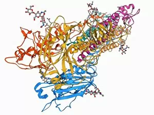
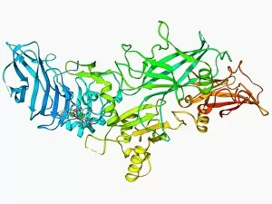
H5N1 Haemagglutinin protein subunit F006 / 9590
H5N1 haemagglutinin protein subunit. Molecular model of the haemagglutinin HA(5) subunit. Haemagglutinin is a surface protein from the influenza A virus

Background imageProteins Collection: Aspartyl-tRNA synthetase protein molecule

Aspartyl-tRNA synthetase protein molecule. Molecular model showing the structure of the active site of aspartyl-tRNA synthetase (DARS) from yeast

Background imageProteins Collection: Transcription factor and DNA molecule F006 / 9484

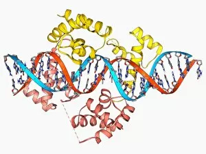
Transcription factor and DNA molecule F006 / 9484
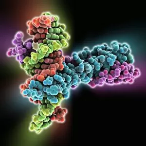
Transcription factor and DNA molecule. Molecular model of glucocorticoid receptor (GR) transcription factor protein (pink and blue) complexed with a molecule of DNA (deoxyribonucleic acid)

Background imageProteins Collection: Haemagglutinin protein subunit F006 / 9479

Haemagglutinin protein subunit F006 / 9479
Haemagglutinin protein subunit. Molecular model of the ectodomain of the haemagglutinin HA(2) subunit. Haemagglutinin is a surface protein from the influenza A virus

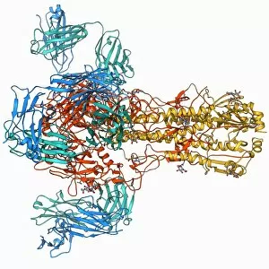
Background imageProteins Collection: Haemagglutinin viral surface protein F006 / 9470

Haemagglutinin viral surface protein F006 / 9470
Haemagglutinin viral surface protein. Molecular model of haemagglutinin, a surface protein from the influenza virus, complexed with a neutralising antibody

Background imageProteins Collection: Human poliovirus, molecular model F006 / 9434

Human poliovirus, molecular model F006 / 9434
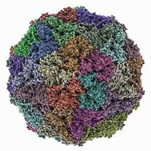
Human poliovirus particle. Molecular model of the capsid of the human poliovirus. The capsid is a protein coat that encloses the viruss genetic information (genome), stored as RNA (ribonucleic acid)

Background imageProteins Collection: VEE equine encephalitis virus capsid

VEE equine encephalitis virus capsid
Venezuelan equine encephalitis virus capsid, molecular model. This mosquito-borne virus can kill horses and other equine species, causing brain and spinal cord inflammation

Background imageProteins Collection: Green fluorescent protein molecule F006 / 9402

Green fluorescent protein molecule F006 / 9402
Green fluorescent protein (GFP), molecular model. The molecule has a cylindrical structure formed from beta sheets (ribbons). GFP is found in the Pacific jellyfish Aequorea victoria

Background imageProteins Collection: Green fluorescent protein molecule F006 / 9343

Green fluorescent protein molecule F006 / 9343
Green fluorescent protein (GFP), molecular model. The molecule has a cylindrical structure formed from beta sheets (ribbons). GFP is found in the Pacific jellyfish Aequorea victoria

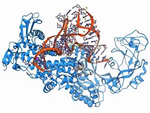
Background imageProteins Collection: Valyl-tRNA synthetase molecule F006 / 9342

Valyl-tRNA synthetase molecule F006 / 9342
Valyl-tRNA synthetase protein molecule. Molecular model showing bacterial valyl-tRNA synthetase complexed with valyl tRNA (transfer ribonucleic acid)

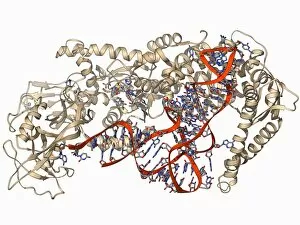
Background imageProteins Collection: Isoleucyl-tRNA synthetase molecule F006 / 9329

Isoleucyl-tRNA synthetase molecule F006 / 9329
Isoleucyl-tRNA synthetase protein molecule. Molecular model showing bacterial isoleucyl-tRNA synthetase complexed with aspartyl tRNA (transfer ribonucleic acid)

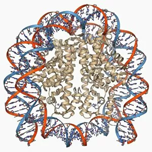
Background imageProteins Collection: Nucleosome molecule F006 / 9323

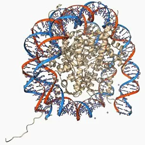
Nucleosome molecule F006 / 9323
Nucleosome, molecular model. A nucleosome is a subunit of chromatin, the substance that forms chromosomes. It consists of a short length of DNA (deoxyribonucleic acid)

Background imageProteins Collection: Nucleosome molecule F006 / 9314

Nucleosome molecule F006 / 9314
Nucleosome, molecular model. A nucleosome is a subunit of chromatin, the substance that forms chromosomes. It consists of a short length of DNA (deoxyribonucleic acid)

Background imageProteins Collection: Green fluorescent protein molecule F006 / 9313

Green fluorescent protein molecule F006 / 9313
Green fluorescent protein (GFP), molecular model. The molecule has a cylindrical structure formed from beta sheets (ribbons). GFP is found in the Pacific jellyfish Aequorea victoria

Background imageProteins Collection: Semliki forest virus capsid F006 / 9297

Semliki forest virus capsid F006 / 9297
Semliki forest virus capsid, molecular model. This virus, named for the forest in Uganda where it was identified, is spread by the bite of mosquitoes. It can infect both humans and animals

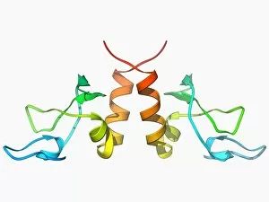
Background imageProteins Collection: HP1 molecule C-terminal domain F006 / 9298

HP1 molecule C-terminal domain F006 / 9298
HP1 molecule C-terminal domain. Molecular model showing the structure of the C terminal (shadow chromo) domain of a heterochromatin protein 1 (HP1) molecule from a mouse

Background imageProteins Collection: Human poliovirus, molecular model F006 / 9289

Human poliovirus, molecular model F006 / 9289
Human poliovirus particle. Molecular model of the capsid of the human poliovirus. The capsid is a protein coat that encloses the viruss genetic information (genome), stored as RNA (ribonucleic acid)

Background imageProteins Collection: MHC protein complexed with flu virus F006 / 9294

MHC protein complexed with flu virus F006 / 9294
MHC protein complexed with flu virus. Molecular model showing human class II MHC (major histocompatibility complex) protein HLA-DR1 complexed with an influenza (flu) virus peptide

Background imageProteins Collection: Yeast DNA recognition, molecular model F006 / 9282

Yeast DNA recognition, molecular model F006 / 9282
Yeast DNA recognition. Computer model showing a GAL4 transcription activator protein bound to a yeast DNA (deoxyribonucleic acid) molecule (red and blue)

Background imageProteins Collection: ATP synthase molecule F006 / 9258

ATP synthase molecule F006 / 9258
ATP synthase molecule. Molecular model showing the structure of ATP synthase (ATPase) subunit A and C. ATPase is an important enzyme that provides energy for cells through the synthesis of adenosine

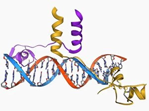
Background imageProteins Collection: Pit-1 transcription factor bound to DNA F006 / 9242

Pit-1 transcription factor bound to DNA F006 / 9242
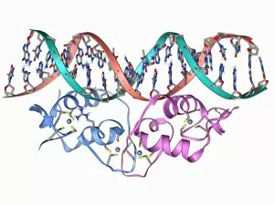
Pit-1 transcription factor bound to DNA. Molecular model showing pituitary-specific positive transcription factor 1 (Pit-1) (yellow and pink) bound to a strand of DNA (deoxyribonucleic acid)

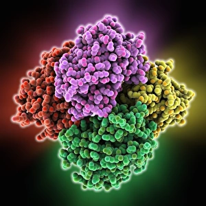
Background imageProteins Collection: Chaperonin protein complex F006 / 9236

Chaperonin protein complex F006 / 9236
Chaperonin protein complex. Molecular model showing the structure of a GroEL GroES (ADP)7 chaperonin complex. Chaperonins are proteins that provide favourable conditions for the correct folding of

Background imageProteins Collection: Aspartyl-tRNA synthetase molecule F006 / 9238

Aspartyl-tRNA synthetase molecule F006 / 9238
Aspartyl-tRNA synthetase protein molecule. Molecular model showing bacterial aspartyl-tRNA synthetase complexed with aspartyl tRNA (transfer ribonucleic acid)

Background imageProteins Collection: Nucleosome molecule F006 / 9235

Nucleosome molecule F006 / 9235
Nucleosome, molecular model. A nucleosome is a subunit of chromatin, the substance that forms chromosomes. It consists of a short length of DNA (deoxyribonucleic acid)

Background imageProteins Collection: Interleukin-6, molecular model F006 / 9232

Interleukin-6, molecular model F006 / 9232
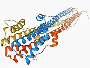
Interleukin-6. Molecular model of the cytokine protein human interleukin-6. This protein is produced in the body and has a wide variety of functions in the immune system

Background imageProteins Collection: Anthrax protective antigen molecule F006 / 9229

Anthrax protective antigen molecule F006 / 9229
Anthrax protective antigen molecule. Computer model showing the structure of a molecule of protective antigen (PA) produced by anthrax (Bacillus anthracis) bacteria

Background imageProteins Collection: Anthrax protective antigen molecule F006 / 9225

Anthrax protective antigen molecule F006 / 9225
Anthrax protective antigen molecule. Computer model showing the structure of a molecule of protective antigen (PA) produced by anthrax (Bacillus anthracis) bacteria

Background imageProteins Collection: Nucleus and endoplasmic reticulum F006 / 9201

Nucleus and endoplasmic reticulum F006 / 9201
Computer artwork showing part of a human or eukaryotic cell. In the middle the nucleus which has a membrane with nuclear pores. Inside the nucleus is the DNA

Background imageProteins Collection: H1N1 flu virus particle, artwork F006 / 9186

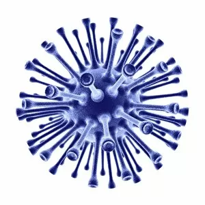
H1N1 flu virus particle, artwork F006 / 9186
H1N1 flu virus particle. Computer artwork of an H1N1 influenza A (flu) virus particle (virion). In the particles lipid envelope (purple) are two types of protein spike

Background imageProteins Collection: Murine minute virus capsid

Murine minute virus capsid, molecular model. This parvovirus infects mice, its only known natural host. It is highly infectious, transmitted through the nose and mouth

Background imageProteins Collection: Zinc finger bound to DNA

Zinc finger bound to DNA. Molecular model showing a zinc finger molecule bound (orange) to a strand of DNA (deoxyribonucleic acid, pink and green)

Background imageProteins Collection: TATA box-binding protein complex C017 / 7089

TATA box-binding protein complex C017 / 7089
TATA box-binding protein complex. Molecular model showing a TATA box-binding protein (TBP, green) complexed with a strand of DNA (deoxyribonucleic acid, spheres) and transcription factor IIB

Background imageProteins Collection: Stem cell-derived astrocyte brain cells

Stem cell-derived astrocyte brain cells
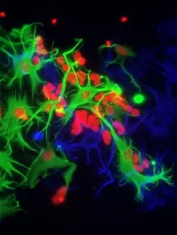
Stem cell-derived nerve cells. Fluorescence light micrograph of astrocyte brain cells that have been derived from neural (nerve) stem cells from a mouse

Background imageProteins Collection: Cell membrane, artwork C018 / 7905

Cell membrane, artwork C018 / 7905
Cell membrane. Computer artwork of a section though a cell membrane. This is a semi-permeable membrane that controls what substances leave and enter the cell

Background imageProteins Collection: SV40 virus capsid, molecular model C018 / 7904

SV40 virus capsid, molecular model C018 / 7904
SV40 virus capsid, molecular model. Simian virus 40 (SV40) is found in monkeys such as Rhesus monkeys and macaques. Potentially tumour-causing, it is used in laboratory research and in vaccines

Background imageProteins Collection: SV40 virus capsid, molecular model C018 / 7903

SV40 virus capsid, molecular model C018 / 7903
SV40 virus capsid, molecular model. Simian virus 40 (SV40) is found in monkeys such as Rhesus monkeys and macaques. Potentially tumour-causing, it is used in laboratory research and in vaccines

Background imageProteins Collection: Antibodies and viruses, artwork

Antibodies and viruses, artwork
Antibodies and viruses. Computer artwork showing antibody, or immunoglobulin, molecules (blue) surrounding virus particles (virions, yellow)

Background imageProteins Collection: T-cell receptor bound to enterotoxin

T-cell receptor bound to enterotoxin, molecular model. The T cell receptor (TCR) is a protein complex found on the surface of a type of white blood cell called T lymphocytes (or T cells)

Background imageProteins Collection: Bird egg white protein, molecular model

Bird egg white protein, molecular model. This is a deglycosylated form of the egg white glycoprotein avidin, obtained from a chicken (Gallus gallus)

Background imageProteins Collection: Reversibly switchable fluorescent protein

Reversibly switchable fluorescent protein, molecular model. Reversibly switchable fluorescent proteins (RSFPs) are proteins that can be repeatedly converted between a fluorescent

Background imageProteins Collection: Excisionase complex with DNA

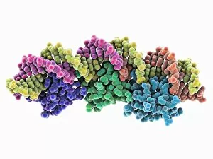
Excisionase complex with DNA. Molecular model of three excisionase proteins (bottom, purple, green and blue) bound to a strand of DNA (top, deoxyribonucleic acid)

Background imageProteins Collection: Epstein-Barr virus protein and DNA

Epstein-Barr virus protein and DNA. Molecular model of the DNA-binding domain of a viral protein (pink-blue) bound to a lytic gene promoter element (viral strand of DNA, left)

Background imageProteins Collection: Antibodies and bacteria, artwork

Antibodies and bacteria, artwork
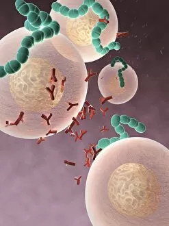
Antibodies and bacteria. Computer artwork showing white blood cells (large, round) and antibody, or immunoglobulin, molecules (red) surrounding bacteria (green)


All Professionally Made to Order for Quick Shipping

Proteins: The Building Blocks of Life From the intricate network of nerve and glial cells to the mesmerizing patterns seen under a light micrograph, proteins play an essential role in every aspect of our existence, and are like the conductors of our body's symphony, orchestrating vital processes that keep us alive and functioning. Take, for example, an anaesthetic inhibiting an ion channel C015 / 6718. Proteins act as gatekeepers, controlling what enters or exits our cells. In this case, they regulate the flow of ions necessary for transmitting nerve signals and maintaining proper cell function. But proteins don't just govern our internal workings; they also interact with external threats such as the avian flu virus. These microscopic invaders hijack host cells using their own protein machinery to replicate themselves. Understanding these interactions is crucial in developing effective treatments against viral infections. While some proteins protect us from harm, others contribute to overall well-being through a balanced diet. Our bodies require various types found in different foods to ensure optimal health and nutrition. The secondary structure is truly a work of art—a complex folding pattern that determines their shape and function. Artists have captured this beauty through stunning artwork showcasing these intricate molecular structures. One such structure is the nucleosome molecule—an elegant arrangement where DNA wraps around protein spools called histones—forming compact units within chromosomes. This organization allows efficient storage and retrieval of genetic information during cell division or gene expression. Antibodies are another remarkable class depicted in captivating artwork. These specialized molecules recognize foreign substances like bacteria or viruses and neutralize them by binding tightly to specific targets on their surface—an extraordinary defense mechanism employed by our immune system. Speaking of bacteria, their ribosomes serve as factories producing new proteins based on instructions encoded in DNA—the blueprint for life itself. Understanding bacterial ribosomes has led to groundbreaking discoveries in antibiotic development, combating infectious diseases that threaten human health.