Fraud Blocker Skip to main content

Proteins Collection (page 8)

Proteins: The Building Blocks of Life From the intricate network of nerve and glial cells to the mesmerizing patterns seen under a light micrograph

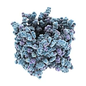
Background imageProteins Collection: Hemolysin-coregulated protein

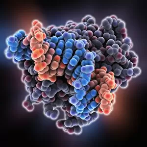
Hemolysin-coregulated protein, molecular model. This protein is from the bacterium Pseudomonas aeruginosa. It is a hexameric ring structure, named hcp1
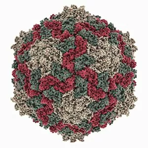
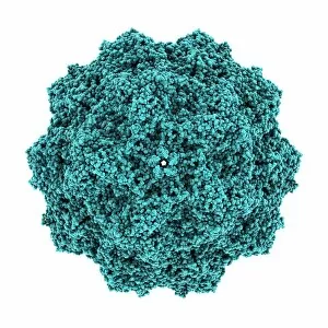
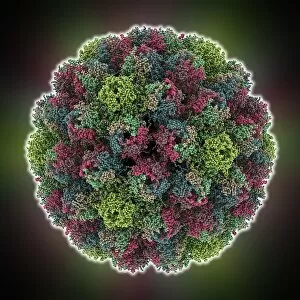
Background imageProteins Collection: Canine parvovirus capsid C015 / 8459

Canine parvovirus capsid C015 / 8459
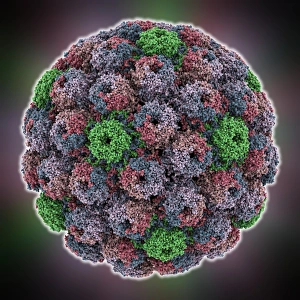
Canine parvovirus capsid, molecular model. Canine parvovirus type 2 (CPV2) was first recognized in 1978 and spread worldwide in less than two years

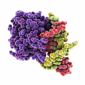
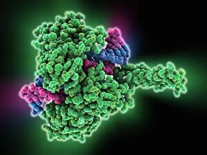
Background imageProteins Collection: DNA repair protein AlkB with DNA C016 / 0546

DNA repair protein AlkB with DNA C016 / 0546
DNA repair protein AlkB with DNA. Molecular model of the DNA (deoxyribonucleic acid) repair protein AlkB (purple) bound to a strand of double-stranded DNA (ds-DNA, red and green)

Background imageProteins Collection: Interferon-DNA transcription complex C015 / 8251

Interferon-DNA transcription complex C015 / 8251
Interferon-DNA transcription complex, molecular model. Bound to the DNA (deoxyribonucleic acid, green and yellow) is transcription factor p65, interferon regulatory factor 7

Background imageProteins Collection: Interferon-DNA transcription complex C015 / 8252

Interferon-DNA transcription complex C015 / 8252
Interferon-DNA transcription complex, molecular model. Bound to the DNA (deoxyribonucleic acid, pink and white) is transcription factor p65, interferon regulatory factor 7, interferon fusion protein

Background imageProteins Collection: Vitamin B12 import proteins C015 / 9942

Vitamin B12 import proteins C015 / 9942
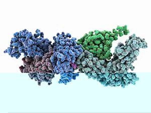
Vitamin B12 import proteins, molecular model. This complex is the import proteins btuC, btuD, and btuF. The first two together form BtuCD

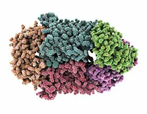
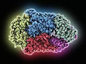
Background imageProteins Collection: Haemoglobin, molecular model C015 / 8939

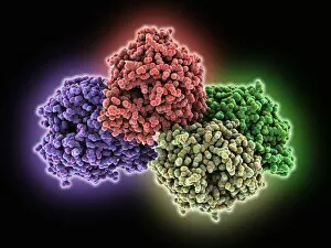
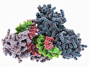
Haemoglobin, molecular model C015 / 8939
Haemoglobin, molecular model. This molecule transports oxygen around the body in red blood cells. It consists of four globin proteins (amino acid chains; orange, green, blue and purple)

Background imageProteins Collection: VDAC-1 ion channel protein C015 / 8249

VDAC-1 ion channel protein C015 / 8249
VDAC-1 ion channel protein, molecular model. This is the human voltage-dependent anion-selective channel protein 1 (VDAC-1)

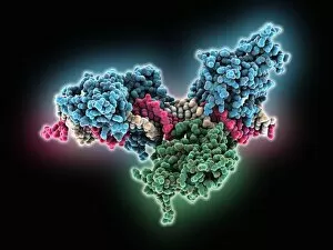
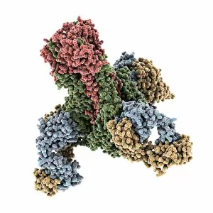
Background imageProteins Collection: Thrombin complexed with fibrinogen C015 / 7149

Thrombin complexed with fibrinogen C015 / 7149
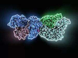
Thrombin complexed with fibrinogen, molecular model. The thrombin molecules (left and right, brown and pink) are bound to the central part of the fibrinogen molecule (centre, multiple colours)

Background imageProteins Collection: Simian virus (SV40) large T antigen C015 / 7069

Simian virus (SV40) large T antigen C015 / 7069
Simian virus (SV40) large T antigen, molecular model. This antigen is from the simian vacuolating virus 40 (SV40). Large T antigens play a role in regulating the viral life cycle of

Background imageProteins Collection: Haemagglutinin viral surface protein C015 / 7123

Haemagglutinin viral surface protein C015 / 7123
Haemagglutinin viral surface protein. Molecular model of haemagglutinin, a surface protein from the influenza virus, complexed with a neutralising antibody

Background imageProteins Collection: 3-hydroxyacyl-CoA dehydrogenase C015 / 9941

3-hydroxyacyl-CoA dehydrogenase C015 / 9941
3-hydroxyacyl-CoA dehydrogenase, molecular model. This enzyme is found in human heart tissue, and catalyzes a reaction that is part of the beta-oxidation pathway

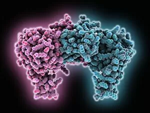
Background imageProteins Collection: Vitamin B12 import proteins C015 / 9943

Vitamin B12 import proteins C015 / 9943
Vitamin B12 import proteins, molecular model. This complex is the import proteins btuC, btuD, and btuF. The first two together form BtuCD

Background imageProteins Collection: Calcium ATPase ion pump C015 / 7142

Calcium ATPase ion pump C015 / 7142
Calcium ATPase ion pump, molecular model. This enzyme is found in muscle cell membranes, where it pumps calcium in and out of muscle cells and controls muscle contractions

Background imageProteins Collection: Retinal-producing oxygenase enzyme C015 / 7151

Retinal-producing oxygenase enzyme C015 / 7151
Retinal-producing oxygenase enzyme, molecular model. This enzyme, an oxygenase, plays a role in the production and metabolism of retinal and other apocarotenoids

Background imageProteins Collection: Adeno-associated virus, molecular model C018 / 0449

Adeno-associated virus, molecular model C018 / 0449
Adeno-associated virus (aV), molecular model. This image shows the core protein of the non-enveloped virus that surrounds the genetic material

Background imageProteins Collection: Hepatitis B virus, molecular model C018 / 0455

Hepatitis B virus, molecular model C018 / 0455
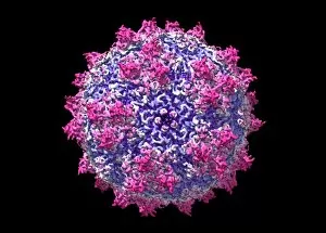
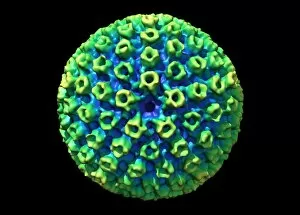
Hepatitis B virus core protein, molecular model. The virus causes hepatitis B, an inflammatory liver disease. The core proteins enclose the virus DNA and are in turn surrounded by a lipid envelope

Background imageProteins Collection: Norovirus capsid, molecular model C018 / 0457

Norovirus capsid, molecular model C018 / 0457
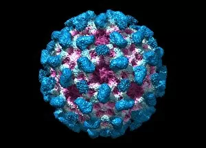
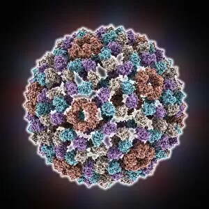
Norovirus capsid, molecular model. Also known as the winter vomiting bug, Noroviruses cause gastroenteritis and are highly contagious, infecting approximately 267 million people a year

Background imageProteins Collection: Astrovirus capsid, molecular model C018 / 0450

Astrovirus capsid, molecular model C018 / 0450
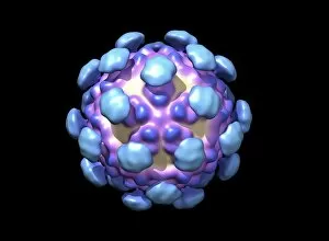
Astrovirus capsid, molecular model. This icosahedral virus was identified in 1975 using electron microscopy. It has a characteristic five-pointed symmetry to its surface, as seen here

Background imageProteins Collection: Hepatitis E virus, molecular model C018 / 0445

Hepatitis E virus, molecular model C018 / 0445
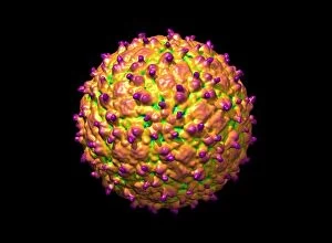
Hepatitis E virus core protein, molecular model. The virus causes hepatitis E, an inflammatory liver disease that usually only lasts a few weeks

Background imageProteins Collection: Human polio virus, molecular model

Human polio virus, molecular model
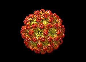
Human polio virus capsid, molecular model. Poliovirus causes poliomyelitis, a disease that can cause paralysis in up to 2 percent of patients, and in some cases death

Background imageProteins Collection: Clathrin lattice, molecular model C018 / 0453

Clathrin lattice, molecular model C018 / 0453
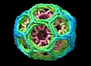
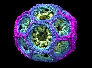
Clathrin lattice, molecular model. This polyhedral protein lattice coats eukaryotic cell membranes (vesicles) and is involved in protein secretion and membrane trafficking

Background imageProteins Collection: Clathrin lattice, molecular model C018 / 0452

Clathrin lattice, molecular model C018 / 0452
Clathrin lattice, molecular model. This polyhedral protein lattice coats eukaryotic cell membranes (vesicles) and is involved in protein secretion and membrane trafficking

Background imageProteins Collection: Clathrin lattice, molecular model C018 / 0454

Clathrin lattice, molecular model C018 / 0454
Clathrin lattice, molecular model. This polyhedral protein lattice coats eukaryotic cell membranes (vesicles) and is involved in protein secretion and membrane trafficking

Background imageProteins Collection: KSHV virus capsid, molecular model C018 / 0456

KSHV virus capsid, molecular model C018 / 0456
KSHV virus capsid, molecular model. KSHV is Kaposis sarcoma-associated herpesvirus. The virus is an oncovirus, which is a virus that can cause cancer

Background imageProteins Collection: Tumour suppressor protein and DNA C017 / 3643

Tumour suppressor protein and DNA C017 / 3643
Tumour suppressor protein and DNA. Computer artwork showing a molecule of the tumour suppressor protein p53 (blue and pink) bound to a molecule of DNA (deoxyribonucleic acid, yellow and orange)

Background imageProteins Collection: Human immune response molecule complex

Human immune response molecule complex. Molecular model showing a human T-cell receptor and an HLA-A leukocyte (white blood cell) antigen bound to a TAX peptide from a virus

Background imageProteins Collection: SV40 virus capsid, molecular model

SV40 virus capsid, molecular model. Simian virus 40 (SV40) is found in monkeys such as Rhesus monkeys and macaques. Potentially tumour-causing, it is used in laboratory research and in vaccines

Background imageProteins Collection: Dengue virus capsid, molecular model

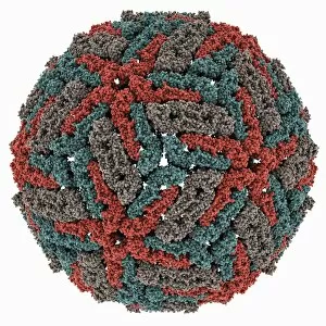
Dengue virus capsid, molecular model. This virus, transmitted by mosquito bites, causes the tropical disease dengue fever in humans

Background imageProteins Collection: Pho4 transcription factor bound to DNA

Pho4 transcription factor bound to DNA. Molecular model showing phosphate system positive regulatory protein (Pho4) (pink and green) bound to a strand of DNA (deoxyribonucleic acid)

Background imageProteins Collection: TATA box-binding protein complex C017 / 7087

TATA box-binding protein complex C017 / 7087
TATA box-binding protein complex. Molecular model showing a TATA box-binding protein (TBP, green) complexed with a strand of DNA (deoxyribonucleic acid, yellow) and transcription factor IIB

Background imageProteins Collection: Human immune response molecule complex C014 / 0871

Human immune response molecule complex C014 / 0871
Human immune response molecule complex. Molecular model showing a human T-cell receptor and an HLA-A leukocyte (white blood cell) antigen bound to a TAX peptide from a virus

Background imageProteins Collection: Type I topoisomerase bound to DNA C014 / 0862

Type I topoisomerase bound to DNA C014 / 0862
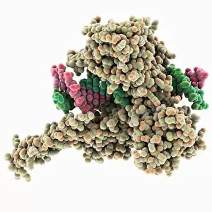
Type I topoisomerase bound to DNA. Molecular model showing a type I topoisomerase molecule (khaki) bound to a strand of DNA (deoxyribonucleic acid, pink and green)

Background imageProteins Collection: Aspartyl-tRNA synthetase protein molecule C014 / 0874

Aspartyl-tRNA synthetase protein molecule C014 / 0874
Aspartyl-tRNA synthetase protein molecule. Molecular model showing the structure of the active site of aspartyl-tRNA synthetase (DARS) from yeast

Background imageProteins Collection: Transcription factor bound to DNA C014 / 0868

Transcription factor bound to DNA C014 / 0868
Transcription factor bound to DNA. Molecular model showing a MATa1/MATalpha2 homeodomain heterodimer (green and pink) in complex with a strand of DNA (deoxyribonucleic acid, orange and blue)

Background imageProteins Collection: Amyloid precursor protein molecule

Amyloid precursor protein molecule
Amyloid precursor protein. Molecular model showing the structure of the protease inhibitor domain of an amyloid precursor protein (APP)

Background imageProteins Collection: Amyloid precursor protein molecule C014 / 0863

Amyloid precursor protein molecule C014 / 0863
Amyloid precursor protein. Molecular model showing the structure of the protease inhibitor domain of an amyloid precursor protein (APP)

Background imageProteins Collection: Ryegrass mottle virus capsid

Ryegrass mottle virus capsid, molecular model. This plant virus is named for its infection of ryegrass, and the discolouration it causes

Background imageProteins Collection: Norwalk virus capsid, molecular model

Norwalk virus capsid, molecular model. This norovirus, which causes a viral form of gastroenteritis, is transmitted from person-to-person or through contaminated food

Background imageProteins Collection: Semliki forest virus capsid

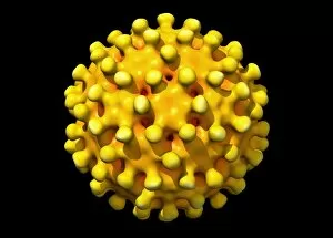
Semliki forest virus capsid, molecular model. This virus, named for the forest in Uganda where it was identified, is spread by the bite of mosquitoes. It can infect both humans and animals

Background imageProteins Collection: TATA box-binding protein complex C014 / 0879

TATA box-binding protein complex C014 / 0879
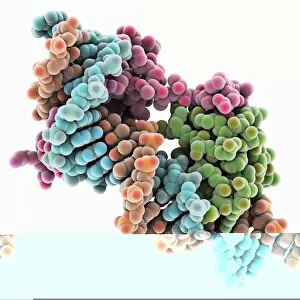
TATA box-binding protein complex. Molecular model showing a TATA box-binding protein (TBP, lilac) complexed with a strand of DNA (deoxyribonucleic acid, green and red)

Background imageProteins Collection: Physalis mottle virus capsid

Physalis mottle virus capsid
Avian polyomavirus capsid, molecular model. This virus, one of a range named for their potential to cause multiple tumours, infects birds. Discovered in budgerigars in 1981, it is often fatal

Background imageProteins Collection: Methyladenine glycosylase bound to DNA C014 / 0877

Methyladenine glycosylase bound to DNA C014 / 0877
Methyladenine glycosylase bound to DNA. Computer model showing a molecule of human DNA-3-methyladenine glycosylase (purple) in complex with DNA (deoxyribonucleic acid, green and orange)

Background imageProteins Collection: Type I topoisomerase bound to DNA C014 / 0883

Type I topoisomerase bound to DNA C014 / 0883
Type I topoisomerase bound to DNA. Molecular model showing a type I topoisomerase molecule (green) bound to a strand of DNA (deoxyribonucleic acid, pink and blue)

Background imageProteins Collection: Bombyx mori densovirus 1 capsid

Bombyx mori densovirus 1 capsid
Bombyx mori densovirus 1 (BmDNV-1), molecular model. This virus infects crustaceans and insects, especially the silkworm (Bombyx mori)

Background imageProteins Collection: Methyladenine glycosylase bound to DNA

Methyladenine glycosylase bound to DNA. Computer model showing a molecule of human DNA-3-methyladenine glycosylase (purple) in complex with DNA (deoxyribonucleic acid, blue and orange)

Background imageProteins Collection: Hepatitis B virus capsid, molcular model

Hepatitis B virus capsid, molcular model
Hepatitis B virus capsid, molecular model. This virus, transmitted through infected bodily fluids or blood, causes the disease hepatitis B in humans, leading to acute liver inflammation

Background imageProteins Collection: Simian rotavirus capsid, molecular model

Simian rotavirus capsid, molecular model. This virus is named for its ability to infect the higher primates (simians). Rotaviruses, transmitted by faecal-oral contact


All Professionally Made to Order for Quick Shipping

Proteins: The Building Blocks of Life From the intricate network of nerve and glial cells to the mesmerizing patterns seen under a light micrograph, proteins play an essential role in every aspect of our existence, and are like the conductors of our body's symphony, orchestrating vital processes that keep us alive and functioning. Take, for example, an anaesthetic inhibiting an ion channel C015 / 6718. Proteins act as gatekeepers, controlling what enters or exits our cells. In this case, they regulate the flow of ions necessary for transmitting nerve signals and maintaining proper cell function. But proteins don't just govern our internal workings; they also interact with external threats such as the avian flu virus. These microscopic invaders hijack host cells using their own protein machinery to replicate themselves. Understanding these interactions is crucial in developing effective treatments against viral infections. While some proteins protect us from harm, others contribute to overall well-being through a balanced diet. Our bodies require various types found in different foods to ensure optimal health and nutrition. The secondary structure is truly a work of art—a complex folding pattern that determines their shape and function. Artists have captured this beauty through stunning artwork showcasing these intricate molecular structures. One such structure is the nucleosome molecule—an elegant arrangement where DNA wraps around protein spools called histones—forming compact units within chromosomes. This organization allows efficient storage and retrieval of genetic information during cell division or gene expression. Antibodies are another remarkable class depicted in captivating artwork. These specialized molecules recognize foreign substances like bacteria or viruses and neutralize them by binding tightly to specific targets on their surface—an extraordinary defense mechanism employed by our immune system. Speaking of bacteria, their ribosomes serve as factories producing new proteins based on instructions encoded in DNA—the blueprint for life itself. Understanding bacterial ribosomes has led to groundbreaking discoveries in antibiotic development, combating infectious diseases that threaten human health.